Where is the sternal angle

Emma Valentine
Published Apr 24, 2026
The sternal angle, which varies around 162 degrees in males, marks the approximate level of the 2nd pair of costal cartilages, which attach to the second ribs, and the level of the intervertebral disc between T4 and T5. In clinical applications, the sternal angle can be palpated at the T4 vertebral level.
Which bones form the sternal angle?
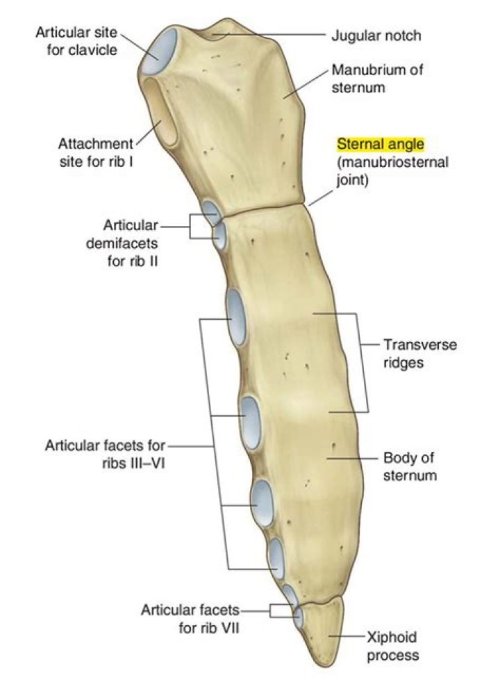
The sternal angle (or manubriosternal joint) is the angle formed (viewed laterally) between the fused manubrium and the corpus sterni. g. The costal notches along either side of the corpus sterni are for articulation with the costal cartilages of ribs 2–7.
What is true sternal angle?
The sternal angle (angle of Louis) is the name of the manubriosternal joint. It is a fibrocartilage joint that allows for some movement acting like a hinge so that the body can move anteriorly during deep inspiration.
How sternal angle is formed?
The sternal angle (of Lewis) is formed by the angle between the manubrium and the body of the sternum at the manubriosternal symphysis (see Fig. 6-2). This angle makes the sternum slightly convex anteriorly. The second costal cartilage articulates with the sternum at this angle.Where does trachea bifurcate sternal angle?
The bifurcation can be located anywhere between the levels of the fourth and seventh thoracic vertebrae. Most commonly it is located at the level of the sternal angle and vertebra T5.
What is the manubrium of the sternum?
The manubrium (manubrium sterni) is quadrangular shaped with four borders. … The clavicular notches of the sternum articulate with the medial end of each clavicle to form the sternoclavicular joints. The manubrium sterni also articulates with the costal cartilages of the 1st pair of ribs.
What is the function of sternum?
Your sternum, along with your ribs, works to protect the organs of your torso, such as your heart, lungs, and chest blood vessels. Support. Your sternum also provides a connection point for other parts of your skeletal system, including your collarbone and most of your ribs.
Where does the sternal angle lie quizlet?
The sternal angle (angle of Louis) is located where and what is it a landmark for? It is located between the manubrium and the body of the sternum– located at the articulation of the 2nd ribs.Why is it called angle of Louis?
1 This anatomic landmark is named after the French surgeon Antoine Louis (1723–1792). Since then, the medical literature has variously described this landmark as the Angle of Louis, Lewis, and Ludwig. … The anatomic verification of this distance comes from relatively few cadaver studies.
What are the events that occur at the sternal angle?- Second costal cartilage and rib lies at this level.
- Superior and inferior mediastinum are demarcated.
- Ascending aorta ends.
- Arch of aorta begins.
- Arch of aorta ends.
- Descending aorta begins.
- Trachea divides into two principal bronchi.
Who discovered the sternal angle of Louis?
The Angle of Louis, more commonly known as the sternal angle or the manubriosternal joint (MSJ), was first described by Pierre Charles Alexandre Louis, a 19 th century Frenchman who postulated that increased angulation was associated with worsening progression of emphysema [1] .
What is thoracic vertebra?
Thoracic vertebrae are the twelve vertebral segments (T1-T12) that make up the thoracic spine. These structures have very little motion because they are firmly attached to the ribs and sternum (breastbone).
Can you feel manubrium?
Xiphoid process The manubrium of the sternum is the superior part of the sternum. The manubrium has the following features: Jugular notch (suprasternal notch) – you can palpate this notch yourself if you feel in the midline between the proximal ends of your clavicles.
What is a costal groove?
costal groove. the groove on the inner surface of the inferior border of the body of the rib. it accommodates the intercostal neurovascular bundle; the costal groove provides a protective function for the intercostal neurovascular bundle, ribs 1-7.
What is anterior to the trachea?
The trachea lies in the superior mediastinum. Anteriorly, it is covered from before backward by the manubrium sterni, the remains of the thymus, the left brachiocephalic (innominate) vein, the aortic arch, the brachiocephalic trunk and left common carotid arteries, and the deep cardiac plexus.
Can you live without a sternum?
Removal of the sternum creates some instability to the rib cage, but most patients do well without an intact sternum. It does, however, create a large space which the overlying skin alone cannot close. The body will fill any such empty space, called dead space, with clotted blood, serum or lymph.
Can you crack your chest bone?
Fractures. A sternum fracture, or break in the breastbone, is usually caused by direct trauma to the bone. The swelling of the joints associated with sternum fractures can cause popping in this area as well.
Why does your sternum hurt?
Costochondritis is the most common cause The most common cause of sternum pain is a condition called costochondritis. This occurs when the cartilage that connects your ribs to your sternum becomes inflamed. Symptoms of costochondritis include: sharp pains or aches on the side of your sternum area.
What level is the manubrium?
In the living, the manubrium is to be found at the level of the third and fourth thoracic vertebrae and forms the anterior boundary of the superior mediastinum.
What does the manubrium look like?
The manubrium is the most superior portion of the sternum. It is trapezoid in shape. The superior aspect of the manubrium is concave, producing a depression known as the jugular notch – this is visible underneath the skin. Either side of the jugular notch, there is a large fossa lined with cartilage.
What is the sternal body?
The sternal body or gladiolus is the middle and largest of the three parts of the sternum. It is formed by the fusion of four sternebrae which finish ossifying after puberty.
How many true ribs do humans have?
In humans there are normally 12 pairs of ribs. The first seven pairs are attached directly to the sternum by costal cartilages and are called true ribs. The 8th, 9th, and 10th pairs—false ribs—do not join the sternum…
What is typical rib?
Typical ribs are those numbered 2 to 10 with ribs 1, 11 and 12 considered atypical. Some authors however include ribs 2 and 10 also atypical.
Where is the false rib?
False rib: One of the last five pairs of ribs. A rib is said to be false if it does not attach to the sternum (the breastbone). The upper three false ribs connect to the costal cartilages of the ribs just above them.
Where is the manubrium bone located?
The manubrium is the superior part of the sternum lying at the level of T3-T4 vertebrae. It forms the superior wall of the anterior mediastinum and its superior border also contributes to the superior thoracic aperture (thoracic inlet).
Which structure separates the thoracic cavity from the abdomen?
The diaphragm is a thin dome-shaped muscle which separates the thoracic cavity (lungs and heart) from the abdominal cavity (intestines, stomach, liver, etc.). It is involved in respiration, drawing downward in the chest on inhalation, and pushing upward in exhalation.
Which part of the respiratory system is referred to as angle of Louis?
Alveoli. Air sac of the lungs. Angle of Louis. Articulation point between the manubrium and the body of the sternum.
How much is Louis angle?
The sternal angle (angle of Louis) is the anterior angle formed by the junction of the manubrium and the body of the sternum which varies around 162 degrees in males.
How do you find the sternal angle of Louis?
To find it on yourself, place your fingers gently at the base of your throat in a central position and move your fingers downward until you can feel the top of the sternum, or rib cage. From this position, continue to move your fingers downward until you feel a boney lump. This is the “angle of Louis”.
Where is T7 located?
The T7 vertebra is the seventh thoracic vertebra, found in the middle of the chest between the seventh and eighth pairs of ribs. It plays important roles in the support of the spinal cord, ribcage, and muscles of the chest.
Where is T5 and T6 in the spine?
T5: Fifth thoracic vertebra. T6: Sixth thoracic vertebra. T7: Seventh thoracic vertebra.