What is short tau inversion recovery

Victoria Simmons
Published Apr 15, 2026
Short tau inversion recovery (STIR), also known as short TI inversion recovery, is a fat suppression technique with an inversion time TI = ln(2)·T1fat, where the signal of fat is zero. This equates to approximately 140 ms at 1.5 T.
What is T2 and stir?
Abstract. T2-weighted short-tau inversion recovery (T2w-STIR) imaging is the best approach for oedema-weighted cardiac magnetic resonance imaging (MRI), as it suppresses the signal from flowing blood and from fat and enhances sensitivity to tissue fluid.
What does high stir signal mean?
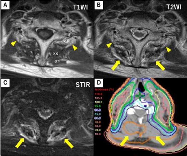
In these MRI images abnormal signal is seen in the vertebral bodies and intervertebral disc. Abnormal low signal on the T1 image and abnormal high signal on the STIR image – indicates abnormal fluid.
Is Stir T1 or T2?
What is STIR? STIR stands for Short-TI Inversion Recovery and is typically used to null the signal from fat. At 1.5T fat has a T1 value of approximately 260 ms, so its TInull value is approximately 0.69 x 250 = 180 ms.What is MRI stir for?
STIR imaging is commonly used to detect bone marrow lesions because it is a sensitive technique for detecting tumor, edema, and infection in bone marrow (15).
What does flair stand for in MRI?
Fluid-attenuated inversion recovery (FLAIR) is an advanced magnetic resonance imaging sequence that reveals tissue T2 prolongation with cerebrospinal fluid suppression, allowing detection of superficial brain lesions.
What are STIR sequences?
The STIR sequence, designed to suppress signal from fat, also enhances the signal from tissue with long T1 and T2 relaxation times, such as neoplastic and inflammatory tissue.
What is hyperintense on STIR MRI?
T1, T2 or FLAIR) to highlight or suppress different types of tissue so that abnormalities can be detected. Hyperintensity on a T2 sequence MRI basically means that the brain tissue in that particular spot differs from the rest of the brain.What does inversion recovery do?
Fluid-attenuated inversion recovery (FLAIR) is an MRI sequence with an inversion recovery set to null fluids. For example, it can be used in brain imaging to suppress cerebrospinal fluid (CSF) effects on the image, so as to bring out the periventricular hyperintense lesions, such as multiple sclerosis (MS) plaques.
What does a high T2 signal mean?An increase in T2 signal intensity is often associated with chronic compression of the spinal cord, and it is well established that chronic compression results in structural changes to the spinal cord.
Article first time published onWhat is T1 and T2 in MRI?
The most common MRI sequences are T1-weighted and T2-weighted scans. T1-weighted images are produced by using short TE and TR times. The contrast and brightness of the image are predominately determined by T1 properties of tissue. Conversely, T2-weighted images are produced by using longer TE and TR times.
What shows up black on an MRI?
MRI can be used to view arteries and veins. Standard MRI can’t see fluid that is moving, such as blood in an artery, and this creates “flow voids” that appear as black holes on the image. Contrast dye (gadolinium) injected into the bloodstream helps the computer “see” the arteries and veins.
What is stir signal abnormality?
When an abnormally bright, diffuse MR signal intensity on STIR imaging is seen more than 6 months after an original injury, such abnormal signal intensity is likely to represent new injury.
What is the difference between stir and T2 fat sat?
STIR sequences have the advantage of increasing the relative signal intensity of fluid as a result of the additive T1 and T2 contrast effect. This allows STIR images to have greater contrast between fluid and other tissues than fat-suppressed T2-weighted fast spin-echo images.
Why is white matter dark on T2?
T2-weighted (T2; long TR and long TE): Water, such as CSF, appears bright, while air appears dark. Fat, such as lipids in the white matter, appears dark.
What is inversion time?
The time between the 180° inverting pulse and the 90°-pulse is called the inversion time (TI). The repetition time (TR) and echo time (TE) are defined as they are for spin echo.
Is white matter the same as lesions?
Those located between the cortex and ventricles, with some space between, are just called “white matter lesions”. There are also subtypes in the “deep white matter”, below the ventricles, some in the cerebellum, and sometimes they are seen in the brainstem.
How long does a FLAIR MRI take?
Notwithstanding very long imaging times (15-20 min typical), the T2-FLAIR technique repeatedly proved itself by revealing a wide range of lesions, including cortical, periventricular, and meningeal diseases that were difficult to see on conventional images.
How do I know if I have T1 or T2 MRI?
- T2 images are a map of proton energy within fatty AND water-based tissues of the body.
- Fatty tissue is distinguished from water-based tissue by comparing with the T1 images – anything that is bright on the T2 images but dark on the T1 images is fluid-based tissue.
What is null point in MRI?
If a tissue happens to have longitudinal magnetization (Mz) close to zero at the end of the TI interval, that tissue will produce little or no signal and make no significant contribution to the magnitude-reconstructed IR image. The tissue is said to be “nulled” or “suppressed”.
What is T2 and flair Hyperintensities?
Focal hyperintensities in the subcortical white matter demonstrated by T2-weighted or FLAIR images are a common incidental finding in patients undergoing brain MRI for indications other than stroke. They are indicative of chronic microvascular disease.
What is mild stir hyperintensity?
White matter hyperintensities (WMHs) are lesions in the brain that show up as areas of increased brightness when visualised by T2-weighted magnetic resonance imaging (MRI). WMH’s are also referred to as Leukoaraiosis and are often found in CT or MRI’s of older patients.
What is hyperintensity of the spine?
Definition. A region of high intensity (brightness) observed upon magnetic resonance imaging (MRI) scans of the spinal cord. [
What is T1 and T2?
T1 and T2 are technical terms applied to different MRI methods used to generate magnetic resonance images. Specifically, T1 and T2 refers to the time taken between magnetic pulses and the image is taken. These different methods are used to detect different structures or chemicals in the central nervous system.
What shows up as white on an MRI?
What Are White Spots? Spots on a brain MRI are caused by changes in water content and fluid movement that occur in brain tissue when the brain cells are inflamed or damaged. These lesions are more easily seen on T2 weighted images, a term that describes the frequency (speed) of the radio impulses used during your scan.
What does a brain MRI without contrast show?
Even without the intravenous contrast, MRI can detect pathology in most organs and in some cases the pathology is made less visible on a contrast MRI than a non-contrast scan. For example, non-contrast scans provide greater images of blood vessel activity to detect aneurysms and blocked blood vessels.
What is dark on T2 MRI?
On a T2-weighted scan compartments filled with water (such as CSF compartments) appear bright and tissues with high fat content (such as white matter) appear dark.
What does T2 signal mean on MRI?
T2 reflects the length of time it takes for the MR signal to decay in the transverse plane. A short T2 means that the signal decays very rapidly. So substances with short T2’s have smaller signals and appear darker than substances with longer T2 values.
What is increased T2 signal on brain MRI report?
A hyperintensity or T2 hyperintensity is an area of high intensity on types of magnetic resonance imaging (MRI) scans of the brain of a human or of another mammal that reflect lesions produced largely by demyelination and axonal loss.
Does CT brain need contrast?
CT of the brain can be done with or without contrast, but it is often not needed. In general, it is preferred that the choice of contrast or no contrast be left up to the discretion of the imaging physician.
Does T2 flair mean MS?
T2 sequences may be used to count the total number of MS lesions or “MS lesion burden.” MS lesions look like white spots on T2 sequences. Fluid attention inversion recovery (FLAIR) sequences are special T2 scans in which signals from the fluid surrounding brain tissue (cerebrospinal fluid or CSF) has been removed.