What is MRI pulse sequence

Victoria Simmons
Published Mar 20, 2026
An MRI pulse sequence is a programmed set of changing magnetic gradients. Each sequence will have a number of parameters, and multiple sequences grouped together into an MRI protocol.
What is pulse sequence MRI?
An MRI pulse sequence is a programmed set of changing magnetic gradients. Each sequence will have a number of parameters, and multiple sequences grouped together into an MRI protocol.
What is pulse sequence diagram?
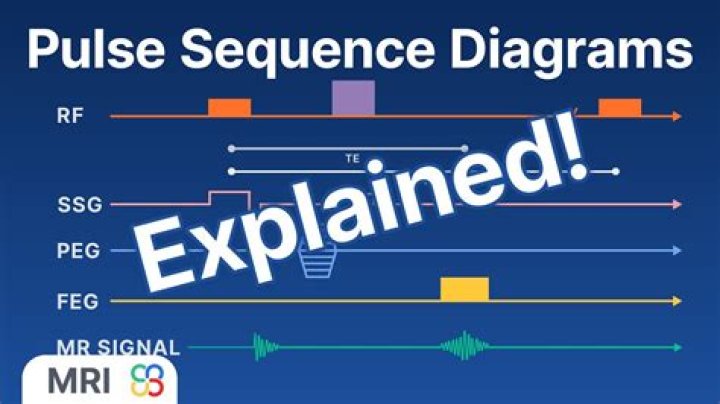
A pulse sequence diagram (PSD) illustrates the sequence of events that occur during magnetic resonance imaging (MRI). It is a timing diagram showing the radio frequency (RF) pulses, gradients, and echoes.
What is the purpose of a pulse sequence?
A pulse sequence is the measurement technique by which an MR image is obtained. As implemented by most manufacturers, the pulse sequence actually executed during the measurement is defined from parameters directly selected by the operator and variables defined in template files.What is pulse NMR?
In the pulsed NMR method, the RF excitation is applied to the sample in a series of short bursts, or pulses. The application of the RF field for a short time (the “pulse width”) allows the applied torque to rotate the net magnetization kM by a specific amount.
How many sequences are there in MRI?
There are two main sequence families, depending on the type of echo recorded: spin echo sequences and gradient echo sequences. There are two main sequence families, depending on the type of echo recorded: spin echo sequences, characterized by the presence of a 180° rephasing RF pulse. gradient echo sequences.
How do you read a MRI sequence?
- Start by checking the patient and image details.
- Look at all the available image planes.
- Compare the fat-sensitive with the water-sensitive images looking for abnormal signal.
- Correlate the MRI appearances with available previous imaging.
- Relate your findings to the clinical question.
What is MRI flair for?
Fluid-attenuated inversion recovery (FLAIR) is an MRI sequence with an inversion recovery set to null fluids. For example, it can be used in brain imaging to suppress cerebrospinal fluid (CSF) effects on the image, so as to bring out the periventricular hyperintense lesions, such as multiple sclerosis (MS) plaques.What are the special sequences for MRI brain?
The most common MRI sequences are T1-weighted and T2-weighted scans. T1-weighted images are produced by using short TE and TR times. The contrast and brightness of the image are predominately determined by T1 properties of tissue. Conversely, T2-weighted images are produced by using longer TE and TR times.
What is spin echo pulse sequence in MRI?The spin echo sequence is made up of a series of events : 90° pulse – 180° rephasing pulse at TE/2 – signal reading at TE. This series is repeated at each time interval TR (Repetition time). With each repetition, a k-space line is filled, thanks to a different phase encoding.
Article first time published onWhat is the fastest MRI sequence?
Echo planar imaging (EPI) is perhaps the fastest sequence available and is the most common sequence used for diffusion-weighted imaging. It essentially forms rapidly alternating gradient echoes within a spin echo sequence.
What is a 90 degree RF pulse?
90 RF pulse rotates the net magnetization vector to transverse plane . 180 RF pulse rotates the net magnetization to –Z direction. RF pulse can disturb the protons and transfer energy only when tuned to the precession frequency of the spinning electrons. This phenomenon is called resonance.
What is pulse angle?
Pulses are described using two parameters, the angle and the phase. Typically, the angle and the phase are shown above the pulse. The angle can vary from 0°-180° While the phase can take on values from 0°-360°. Commonly the phase of the pulse is referred to the axis along which the B1 field is applied.
What is a PI 2 pulse?
π2 pulse means that all the particles in the system have gone to the higher level. π pulse excites all particles in the first half time and de-exites in the second, so all particles are in lower level.
Why are RF pulses used in NMR?
With NMR, a radiofrequency (RF) pulse is applied which is able to “tip the spins” so that the direction of the macroscopic magnetization moves into the XY plane and, thus, is able to produce Page 2 Pulse sequence Pavlicek, et at. 50 RadioGraphics January 1984 Volume 4, Special Edition a signal in the detector.
What is T1 and T2?
T1 and T2 are technical terms applied to different MRI methods used to generate magnetic resonance images. Specifically, T1 and T2 refers to the time taken between magnetic pulses and the image is taken. These different methods are used to detect different structures or chemicals in the central nervous system.
What is T1 and T2 relaxation time?
The approach of the system to thermal equilibrium is known as relaxation and T1 and T2 are relaxation times (relaxation rates R1(2)=1/T1(2) are also used). Both relaxation times are time constants used to characterize what are assumed to be first order rate processes.
What does high T2 signal mean on MRI?
An increase in T2 signal intensity is often associated with chronic compression of the spinal cord, and it is well established that chronic compression results in structural changes to the spinal cord.
What does Leukoaraiosis mean?
Background— Leukoaraiosis, a term that defines an abnormal appearance of the subcortical white matter of the brain on neuroimaging (bilateral patchy or diffuse areas of low attenuation on CT or hyperintense T2 MR areas), has gained evidence in retrospective studies to demonstrate its association with stroke and in …
What is T2 and flair signal?
T2/FLAIR. T2/FLAIR images show the total amount of scar from MS from its onset. The pictures show both old and new inflammation. T2/FLAIR lesions can directly account for some symptoms. For example, a brainstem lesion can cause room spinning sensations and balance problems.
What is T2 flair sequence in MRI?
FLAIR MRI is a heavily T2-weighted technique that dampens the ventricular (ie, free-water) CSF signal. Thus, the highest signals on the sequence are from certain brain parenchymal abnormalities, such as MS lesions, while the CSF appears black.
What are the types of spin echo pulse sequence?
The two variables of interest in spin echo sequences are the repetition time (TR) and the echo time (TE). All spin echo sequences include a slice selective 90-degree pulse followed by one or more 180 degree refocusing pulses as shown in the diagrams.
What is GRE sequence?
Gradient echo sequences (GRE) are an alternative technique to spin-echo sequences, differing from it in two principal points: utilization of gradient fields to generate transverse magnetization. flip angles of less than 90°
What is the difference between T2 and T2 *?
T2* can be considered an “observed” or “effective” T2, whereas the first T2 can be considered the “natural” or “true” T2 of the tissue being imaged. T2* is always less than or equal to T2. T2* results principally from inhomogeneities in the main magnetic field.
What is turbo factor in MRI?
The number of echoes received in the same repetition (during TR time) is called the Turbo Factor or Echo Train Length (ETL).
What is B value in DWI?
Conclusion: In DWI, the optimal b value is 600 s/mm(2) ; multiple b values of 600 s/mm(2) and higher are recommended to differentiate between benign and malignant abdominal lesions. The lesion ADC/normal parenchyma ADC ratio is more accurate than using lesion ADC only.
What is haste sequence?
HASTE is an echo-planar fast spin echo sequence trademarked by Siemens. The expanded acronym fairly completely describes what it entails: Half-Fourier Acquisition Single-shot Turbo spin Echo imaging.
What is T1 relaxation in MRI?
The T1 relaxation time, also known as the spin-lattice relaxation time, is a measure of how quickly the net magnetization vector (NMV) recovers to its ground state in the direction of B0.
In which direction is the RF pulse applied?
An rf pulse applies a torque to the bulk magnetisation vector and drives it towards the x–y plane from equilibrium. θ is the pulse tip or flip angle which is most frequently 90 or 180 degrees.
What is T2 relaxation time in MRI?
T2 Relaxation: Definition Thus T2 is the time required for the transverse magnetization to fall to approximately 37% (1/e) of its initial value. Synonyms for T2 relaxation are transverse relaxation and spin-spin relaxation.
What is pulse width NMR?
Pulse width is the length of time that radiofrequency (RF) energy irradiates a sample in nuclear magnetic resonance (NMR) spectroscopy. … The most important part is choosing the length of time for an RF pulse that produces the maximum signal.