What is Mammillary process

Rachel Hickman
Published Mar 19, 2026
The mammillary process is a superior process on costal process
What is the function of Mammillary process?
The primary function associated with the mammillary bodies is recollective memory. Memory information begins within the hippocampus. Theta waves activate CA3 neurons in the hippocampus.
What is mammillary process of lumbar vertebra?
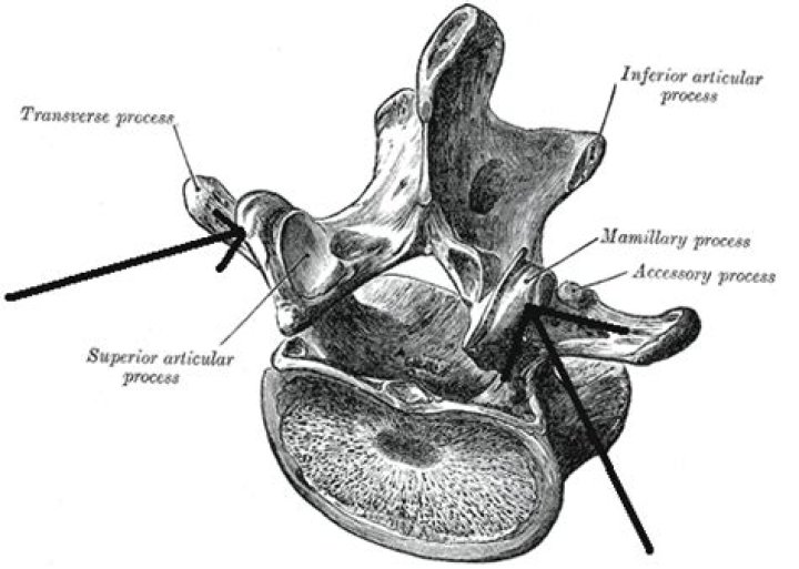
The mammillary process is a somewhat elongated tubercle that originates from the posterolateral margin of the superior articular process. g. The accessory process is a variable, diminutive tubercle on the dorsal aspect of the base of the lumbar transverse process.
Where is the mammillary process?
The mamillary processes are located between transverse and cranial articular process on thoracic and lumbar vertebrae.What vertebrae have Mammillary processes?
Lumbar vertebrae have mammillary bodies on the superior articular processes not present in thoracic vertebrae. This study observed that TLTV have irregular mammillary bodies present in both the thoracic and lumbar regions.
What are the major functions of the intervertebral discs?
The intervertebral discs have the following functions: They provide cushioning for the vertebrae and reduce the stress caused by impact. By keeping the vertebrae separated from each other, they act as a type of shock absorber for the spine. They help protect the nerves that run down the spine and between the vertebrae.
What attaches to Mammillary process?
The superior, or upper tubercle is the mammillary process which connects with the superior articular process. The multifidus muscle attaches to the mammillary process and this muscle extends through the length of the vertebral column, giving support.
What is intervertebral foramen?
Abstract. The intervertebral foramen serves as the doorway between the spinal canal and periphery. It lies between the pedicles of neighboring vertebrae at all levels in the spine.Is lumbar spondylosis arthritis?
Technically, spondylosis is a form of arthritis—spinal osteoarthritis (osteoarthritis is the most common type of arthritis) to be exact. We tend to think of arthritis as something you get in your hands and knees, but the spine, and all of its bones and joints, can fall victim to its grip as well.
Which vertebrae have accessory and mammillary process?Three portions or tubercles can be noticed in a transverse process of a lower lumbar vertebrae: the lateral or costiform process, the mammillary process, and the accessory process. The costiform is lateral, the mammillary is superior (cranial), and the accessory is inferior (caudal).
Article first time published onWhat is Flavum?
One of a series of bands of elastic tissue that runs between the lamina from the axis to the sacrum, the ligamentum flavum connects the laminae and fuses with the facet joint capsules. … As we age, the ligament loses elastin, and this allows the ligament to encroach on the canal.
What muscles are attached to the lumbar spine?
Lumbar vertebrae provide attachment points for numerous muscles: erector spinae, interspinales, intertransversarii, latissimus dorsi, rotatores, and serratus posterior inferior.
What attaches to the accessory process of the lumbar vertebrae?
It gives attachment to the multifidus and to the medial intertransverse muscle. The accessory process varies in prominence and may be difficult to identify. It gives attachment to the medial intertransverse muscle. The costal element is incorporated in the transverse process (fig.
Is your neck connected to your spine?
The neck is connected to the upper back through a series of seven vertebral segments. The cervical spine has 7 stacked bones called vertebrae, labeled C1 through C7. The top of the cervical spine connects to the skull, and the bottom connects to the upper back at about shoulder level.
How are vertebrae connected?
Each vertebra is held to the others by groups of ligaments. Ligaments connect bones to bones; tendons connect muscles to bones. There are also tendons that fasten muscles to the vertebrae. The spinal column also has real joints (just like the knee or elbow or any other joints) called facet joints.
What is the connection between the vertebrae bodies and arches the type of connection?
Adjacent vertebral arches are connected by synovial joints called zygapophysial (facet) joints. They are formed between superior and inferior articular facets. These joints facilitate flexion and extension in the cervical and thoracic spines.
What type of joint is intervertebral joint?
TypeIntervertebral disc joint: Cartilaginous joint; symphysis Zygapophyseal joint: Synovial plane joint, nonaxial, uniplanarLigamentsLongitudinal ligaments (anterior, posterior), ligamenta flava, interspinous ligaments, intertransverse ligaments, supraspinous ligaments, nuchal ligament (cervical spine only)
What are the functions of the vertebral and intervertebral foramina?
The vertebral foramen provides for passage of the spinal cord. Each spinal nerve exits through an intervertebral foramen, located between adjacent vertebrae. Intervertebral discs unite the bodies of adjacent vertebrae.
What are transverse processes?
Transverse process is a small bony projection off the right and left side of each vertebrae. The two transverse processes of each vertebrae function as the site of attachment for muscles and ligaments of the spine as well as the point of articulation of the ribs (in the thoracic spine).
What causes intervertebral disc degeneration?
Underlying causes of disc degeneration include genetic inheritance, age, inadequate metabolite transport, and loading history, all of which can weaken discs to such an extent that structural failure occurs during the activities of daily living.
Why is an intervertebral disc not present between C1 and C2?
C1 and C2 are very specialized vertebrae hence why they don’t have an intervertebral disc. C1 is known as atlas and C2 is known as axis and together, these bones make a pivot joint. … C1 will allow for anterior posterior movements through the dens and C2 will allow for lateral movements.
How do intervertebral discs get nutrients?
The disc is avascular, and the disc cells depend on diffusion from blood vessels at the disc’s margins to supply the nutrients essential for cellular activity and viability and to remove metabolic wastes such as lactic acid.
What should be avoided in lumbar spondylosis?
There should be restriction of heavy lifting, excessive bending, twisting or stooping and avoidance of any work or recreational activities that causes stress to the lumbar spine.
Is walking good for lumbar spondylosis?
Walking strengthens the muscles that support your spine Your trunk, core, and lumbar (lower back) muscles play a vital role in maintaining the stability and movement of your lower back. These muscles can become deconditioned and weak from a sedentary lifestyle, causing malalignment of the spine.
What is the most serious complication of spondylosis?
The main complication of spondylosis is low back, mid back, or neck pain. Usually the back and neck pain caused by spondylosis is not serious, but some people develop chronic pain due to their condition. It is unusual for spondylosis to cause serious neurologic dysfunction due to nerve compression.
What is spiral cord?
A column of nerve tissue that runs from the base of the skull down the center of the back. It is covered by three thin layers of protective tissue called membranes. The spinal cord and membranes are surrounded by the vertebrae (back bones).
What is Atlas vertebra?
atlas: the first cervical vertebra (C1), lying directly under the skull, through which the head articulates with the neck. The main connection to the vertebra below is a pivot around the odontoid process that is an upward projection of the body of the second cervical vertebra.
What exits the intervertebral foramen?
The intervertebral foramina are essentially “exit routes” from which the nerve roots leave the spine and branch out to all parts of the body. Without the foramen, nerve signals could not travel to and from the brain to the rest of the body.
Where is the intervertebral disc?
An intervertebral disc (or intervertebral fibrocartilage) lies between adjacent vertebrae in the vertebral column. Each disc forms a fibrocartilaginous joint (a symphysis), to allow slight movement of the vertebrae, to act as a ligament to hold the vertebrae together, and to function as a shock absorber for the spine.
What kind of tissue makes up the intervertebral discs?
Fibrocartilage is the tough, very strong tissue found predominantly in the intervertebral disks and at the insertions of ligaments and tendons; it is similar to other fibrous tissues but contains cartilage ground substance and chondrocytes.
Is the lumbar vertebrae medial or lateral?
The main distinguishing feature of the lumbar vertebrae is the orientation of the facets on the superior and inferior articular processes. The facets on the superior articular processes face medially and posteriorly, and the facets on the inferior articular processes face laterally and anteriorly.