What is lateral Trochlear

Christopher Lucas
Published Feb 17, 2026
Lateral trochlear inclination (LTI) is the inclination angle between the femoral trochlea and a posterior condylar tangential line 1.
Where is the trochlea located in the knee?
The trochlea is a groove in the femur bone underneath the kneecap (patella). The walls of the trochlea stabilize the patella and allow it to glide down the center of the trochlea as the knee bends.
Is the Trochlear notch medial or lateral?
Trochlear notchFMA23619Anatomical terms of bone
What is trochlea of femur?
Description. The trochlea of femur (femoral trochlea) is the cranial cartilaginous part of distal femur, for articulation with the patella fo form the femoral patellar joint. It consists of a groove bounded by the medial and a lateral ridges.What is Trochlear?
Definition of trochlea : an anatomical structure that is held to resemble a pulley especially : the articular surface on the medial condyle of the humerus that articulates with the ulna.
Where is kneecap located?
The patella (kneecap) is located at the front of the knee joint, within the patellofemoral groove of the femur.
Where is the Trochlear groove?
When the knee is bent, the undersurface of the kneecap (the patella) lies in an area known as the trochlear groove. The sides of the patella and the walls of the groove should be almost parallel. The normal shape of the trochlea groove is concave.
What bones have a trochlea?
The trochlea is the roughly hourglass-shaped feature on the distal end of the humerus. It articulates with the trochlear notch of the ulna.What side is the ulna on?
The forearm consists of two bones, the radius and the ulna, with the ulna is located on the pinky side and the radius on your thumb side.
What is trochlea of Talus?The trochlea of talus is a convex articular surface for the proximal part of articulation with tibia and fibula (only with tibia in horses). The trochlea presents a sagittal groove that articulates with the sagittal ridge of cochlea of tibia.
Article first time published onWhat is the central trochlea?
Answer: The trochlea is considered part of the patellofemoral compartment, Stout says. “ The trochlea is the depression on the anterior distal femur where the patella articulates with the femur,” she says. “
Where is the adductor tubercle?
The adductor tubercle is a bony protuberance on the medial condyle of the femur and is located superior to the medial epicondyle. It demarcates the inferior most aspect of the medial supracondylar line.
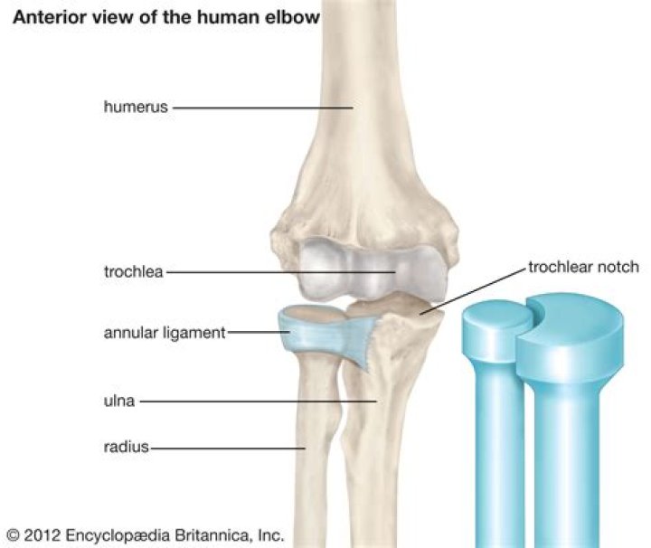
What sits in the trochlear notch?
The proximal end of the ulna resembles a crescent wrench with its large, C-shaped trochlear notch. This region articulates with the trochlea of the humerus as part of the elbow joint. The inferior margin of the trochlear notch is formed by a prominent lip of bone called the coronoid process of the ulna.
What makes up the trochlear notch?
The trochlear notch (semilunar notch, greater sigmoid cavity) is a large depression, formed by the olecranon and the coronoid process, and serving for articulation with the trochlea of the humerus.
Is the trochlea anterior or posterior?
The medial portion of the articular surface of distal humerus is named the trochlea, and presents a deep depression between two well-marked borders; it is convex from before backward, concave from side to side, and occupies the anterior, lower, and posterior parts of the extremity.
Where is the trochlear nerve located?
The Trochlear Nerve (IV) The fourth nerve nucleus is located in the tegmentum of the midbrain at the level of the inferior colliculus, ventral to the periaqueductal gray matter, inferior to the oculomotor nucleus, and superior to the medial longitudinal bundle.
Where is the trochlear nerve in the brain?
The trochlear nerve emerges from the back (dorsal) brainstem, just below the inferior colliculus. It circles from behind around the brainstem and runs forward toward the eye in the subarachnoid space.
How common is Trochlear nerve palsy?
The annual incidence of trochlear nerve palsy was found to be 5.73 per 100,000 per year. [2] In several studies, it has been observed that this entity is much more common in the male gender. [5] This may be due to a higher incidence of head trauma in males.
Is Trochlear dysplasia congenital?
Trochlear dysplasia is characterized by abnormal trochlear morphology and a shallow groove. It is associated with recurrent patellar dislocation, but it is unclear whether the dysplasia is congenital, the result of lateral tracking and chronic instability, or caused by a combination of factors.
What does patellofemoral arthritis feel like?
Patients experiencing patellofemoral knee arthritis will have kneecap pain and stiffness and often swelling in the front part of the knee that typically worsens when walking on inclined terrain, going up and down stairs, squatting or rising from a seated position.
What is the best treatment for chondromalacia patella?
- Placing of an ice or cold pack to the area for 15-20 minutes, four times daily, for several days. …
- Nonsteroidal anti-inflammatory drugs (NSAIDs) for pain relief—These include ibuprofen, naproxen, and aspirin.
What is the outside of your knee called?
The outside half (farthest away from the other knee) is called the lateral tibial plateau, and the inside half (closest to the other knee) is called the medial tibial plateau. The patella glides through a special groove formed by the two femoral condyles called the patellofemoral groove.
What does it mean when the back of your leg hurts behind the knee?
Some of the most common causes of pain behind the knee (posterior knee pain) include, Baker’s cyst, arthritis, infection, injury, tumor, or deep vein thrombosis. Since the knee is the largest and most complex joint in the body, it makes sense that it might hurt sometimes.
What is the part below knee called?
The leg from the knee to the ankle is called the crus or cnemis /ˈniːmɪs/. The calf is the back portion, and the tibia or shinbone together with the smaller fibula make up the front of the lower leg.
Is the ulna medial or lateral?
The ulna is a long bone found in the forearm that stretches from the elbow to the smallest finger, and when in anatomical position, is found on the medial side of the forearm. It is broader close to the elbow, and narrows as it approaches the wrist.
How do you tell the difference between ulna and radius?
The radius connects to the thumb side of your wrist and is the larger of the two while the ulna connects to the pinky side and is the smaller one. An easy way to remember the difference between the two is the word radius is longer than the word ulna just like the bones themselves.
Where is the lateral epicondyle of the humerus?
There are bony bumps at the bottom of the humerus called epicondyles, where several muscles of the forearm begin their course. The bony bump on the outside (lateral side) of the elbow is called the lateral epicondyle.
What is the lateral bone of the forearm?
RadiusTA21210FMA23463Anatomical terms of bone
What is the function of the lateral epicondyle?
Anatomical terms of bone The lateral epicondyle of the humerus is a large, tuberculated eminence, curved a little forward, and giving attachment to the radial collateral ligament of the elbow joint, and to a tendon common to the origin of the supinator and some of the extensor muscles.
Where can I find trochlea of talus?
The trochlea is broader in front than behind, convex from before backward, slightly concave from side to side: in front it is continuous with the upper surface of the neck of the bone.
What bone has a lateral malleolus?
Lateral Malleolus: Bony protrusion felt on the outside of the ankle. The lateral Malleolus is the low end of the Fibula.