What is cell wall staining

William Taylor
Published Mar 18, 2026
Cell staining is a technique that can be used to better visualize cells and cell components under a microscope. By using different stains, one can preferentially stain certain cell components, such as a nucleus or a cell wall, or the entire cell.
Which is cell wall staining method?
- Step 1: Slide preparation. 1.1 Take a clean glass slide and wash it with soap and water thoroughly so as to remove grease and dirt from it. …
- Step 2: Slide labelling. …
- Step 3: Smear preparation and heat fixation. …
- Step 4: Staining. …
- Step 5: Mounting of slide.
What is used to stain bacterial cell walls?
Cells are stained with crystal violet dye. … This complex is a larger molecule than the original crystal violet stain and iodine and is insoluble in water. A decolorizer such as ethyl alcohol or acetone is added to the sample, which dehydrates the peptidoglycan layer, shrinking and tightening it.
What is the purpose of staining?
The purpose of staining is to increase the contrast between the organisms and the background so that they are more readily seen in the light microscope.Why is cell wall important in Gram staining?
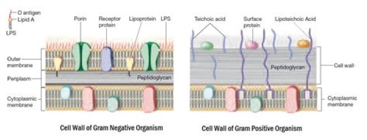
The basic principle of gram staining involves the ability of the bacterial cell wall to retain the crystal violet dye during solvent treatment. Gram-positive microorganisms have higher peptidoglycan content, whereas gram-negative organisms have higher lipid content.
What is the function of cell wall?
The cell wall is the protective, semi-permeable outer layer of a plant cell. A major function of the cell wall is to give the cell strength and structure, and to filter molecules that pass in and out of the cell.
What does an Endospore stain tell you?
Endospore staining is a technique used in bacteriology to identify the presence of endospores in a bacterial sample. Within bacteria, endospores are protective structures used to survive extreme conditions, including high temperatures making them highly resistant to chemicals.
What is staining and its types?
The types are: 1. Simple Staining 2. … Gram Staining 4. Acid Fast Staining 5. Endospore Staining.Why are bacterial cells stained?
Bacteria are stained for better visual observation, to highlight differences, to enhance cell components, to help identify the bacterium, etc.
What are the types of stains?- Oil Stain. Oil stains are the most widely available and the type of stain most people think of when they think of stain. …
- Varnish Stain. Varnish stains resemble oil stains in every way but one. …
- Gel Stain. …
- Lacquer Stain. …
- Water-Soluble Dye Stain. …
- Metal-Complex (Metalized) Dye Stain.
Why staining is important in microbiology?
Cell staining is important in the diagnosis of microorganisms because bacteria can be identified by the color differentiation of stains (dyes). … This staining test highlights differences in the structure of the cell wall of the two types of bacteria.
What is the importance of stain in microbiology?
The most basic reason that cells are stained is to enhance visualization of the cell or certain cellular components under a microscope. Cells may also be stained to highlight metabolic processes or to differentiate between live and dead cells in a sample.
What is staining technique?
Staining is a technique used to enhance contrast in samples, generally at the microscopic level. … Light microscopes are used for viewing stained samples at high magnification, typically using bright-field or epi-fluorescence illumination.
What is difference between gram positive and negative?
Gram positive bacteria have a thick peptidoglycan layer and no outer lipid membrane whilst Gram negative bacteria have a thin peptidoglycan layer and have an outer lipid membrane.
Why Iodine is used in Gram staining?
Gram’s iodine is used in Gram staining procedure to differentiate gram positive and gram negative organisms. Gram’s iodine acts as a mordant that causes the crystal violet to penetrate and adhere to the gram –positive organisms.
What is called cellulose?
Cellulose is a molecule, consisting of hundreds – and sometimes even thousands – of carbon, hydrogen and oxygen atoms. Cellulose is the main substance in the walls of plant cells, helping plants to remain stiff and upright. Humans cannot digest cellulose, but it is important in the diet as fibre.
What are the 3 objectives of the Endospore stain?
The main purpose of endospore staining is to differentiate bacterial spores from other vegetative cells and to differentiate spore formers from non-spore formers.
Why do we use Endospore stain?
Endospore Staining is a technique used in bacteriology to identify the presence of endospores in a bacterial sample, which can be useful for classifying bacteria.
Is Endospore stain positive or negative?
Endospore Stain Only a few genera of bacteria produce endospores and nearly all of them are Gram-positive bacilli.
What is cell and function?
Cells are the basic building blocks of all living things. The human body is composed of trillions of cells. They provide structure for the body, take in nutrients from food, convert those nutrients into energy, and carry out specialized functions.
What is composition of cell wall?
The cell wall is composed of a network of cellulose microfibrils and cross-linking glycans embedded in a highly cross-linked matrix of pectin polysaccharides. In secondary cell walls, lignin may be deposited.
What happens if the cell wall is damaged?
Damage to the cell wall disturbs the state of cell electrolytes, which can activate death pathways (apoptosis or programmed cell death). … A bacterial cell with a damaged cell wall cannot undergo binary fission and is thus certain to die.
What are the advantages of using stains?
The advantage of using stains to look at cells is that stains reveal these details and more. Abnormally shaped or abnormally arranged cells will be evidence of disease. Multiple stains can be simultaneously used on a tissue, such that different cell types appear in different colors.
What is basic staining?
Basic stains, such as methylene blue, Gram safranin, or Gram crystal violet are useful for staining most bacteria. These stains will readily give up a hydroxide ion or accept a hydrogen ion, which leaves the stain positively charged.
What is stain chemical?
Rather than dying the fibers (as with aniline dyes) or putting fine pigment particles on the surface (as with conventional “stains”) a chemical stain reacts with the natural tannin in the wood to produce a brown to reddish brown. The depth of the color that can be achieved is stunning.
What is a stain in microbiology?
staining. [stān´ing] artificial coloration of a substance to facilitate examination of tissues, microorganisms, or other cells under the microscope.
Do basic stains stain the cell?
Because cells typically have negatively charged cell walls, the positive chromophores in basic dyes tend to stick to the cell walls, making them positive stains. Thus, commonly used basic dyes such as basic fuchsin, crystal violet, malachite green, methylene blue, and safranin typically serve as positive stains.
What is the source of stain?
A stain is a discoloration that can be clearly distinguished from the surface, material, or medium it is found upon. They are caused by the chemical or physical interaction of two dissimilar materials. Accidental staining may make materials appear used, degraded or permanently unclean.
How do you identify a stain?
Three criteria for identifying and classifying the most commonly known types of stains are type of edge, feel and colour. All stains cannot be recognized by the appearance of the edge. Here we distinguish between hard and soft stains. Hard stains are caused by Varnish, oil paints and glues.
What is gram negative cell wall?
The Gram-negative cell wall is composed of a thin, inner layer of peptidoglycan and an outer membrane consisting of molecules of phospholipids, lipopolysaccharides (LPS), lipoproteins and sutface proteins. The lipopolysaccharide consists of lipid A and O polysaccharide.
Which antibiotics are best for gram negative bacteria?
- Aminoglycosides (gentamicin, amikacin)
- Glycylcycline (tigecycline)
- Tetracyclines (doxycycline, minocycline)
- Chloramphenicol.
- Sulphonamides (co-trimoxazole)
- Fosfomycin.