What is a localizer in MRI

Mia Morrison
Published Mar 17, 2026
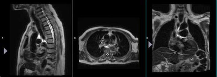
Localizer scans. A set of three-plane, low-resolution, large field-of view localizers are first obtained, equivalent to “scout views” in CT. These localizer images will be used for plotting slices in step 6. 5.
What is a localizer scan?
Localizer images, also called scout images, are used in MR and CT studies to identify the relative anatomical position of a collection of cross-sectional images. A localizer can be acquired as a separate image, as is often done for CTs, or it can be dynamically generated, as is done for MRs.
What are the 3 plane localizers in MRI?
This coordinate system consists of three planes to describe the standard anatomical position of a human. The basic orientation terms for a MRI of the body taken: From the side would be a sagittal plane; from the front, would be a coronal plane; and from the top down, would be a transverse plane.
How does a claustrophobic person get an MRI?
Instead of a tube, an open MRI has scanners on the sides with an opening on top, making it an outstanding option for those who have claustrophobia. The patient lays comfortably on a platform while the scanners on the sides do all the work.What are MRI scout images?
MRI scout images, which are taken prior to a study to determine the range of subsequent images, can be used to rapidly screen the whole brain. We examined whether MRI scout imaging can detect ICHs observed by computed tomography (CT).
What is the Hounsfield unit for blood?
Hounsfield Unit Uncoagulated blood typically measures 30 to 45 HU. Clotted (or concentrated) blood measures higher at 60 to 100 HU. Separated serum plasma is closer to water at 0 to 20 HU. Finally, ascites also has a Hounsfield measurement of around 0 to 20 HU.
Which is an advantage of step and shoot scanning?
Advantage of Axial Scanning In evaluation image quality using phantoms- ‘that do not breathe or move’, step and shoot method result in the highest image quality, superior to that of helical methods.
Which MRI is best for claustrophobia?
The open, upright MRI machine is much better tolerated by patients who are claustrophobic. The scans can be performed with the patient sitting, lying down, or standing.How do you survive a MRI if you are claustrophobic?
- 1-Ask questions beforehand. The more educated and informed you are on the specifics of the test, the less likely you are to be surprised by something. …
- 2-Listen to music. …
- 3-Cover your eyes. …
- 4-Breathe and meditate. …
- 5-Ask for a blanket. …
- 6-Stretch beforehand. …
- 7-Take medication.
If you experience more severe claustrophobia-related symptoms, your doctor may instead recommend intravenous sedation. It’s common to use a combination of Versed (a benzodiazepine) and Fentanyl, an opioid medication commonly prescribed for pain and sedation.
Article first time published onWhat is a 3 plane localizer?
Localizer – a 3-plane localizer or ‘scout’ scan meant to find the subject’s head. It is also be used for prescription for the subsequent scans. Doing some sort of localizer is necessary, and the ‘3planeloc SSFSE’ (single shot fast spin echo) is the standard work-horse used by most CNI users.
What is T1 and T2 in MRI?
The most common MRI sequences are T1-weighted and T2-weighted scans. T1-weighted images are produced by using short TE and TR times. The contrast and brightness of the image are predominately determined by T1 properties of tissue. Conversely, T2-weighted images are produced by using longer TE and TR times.
What is T1-weighted image in MRI?
Definition. A T1-weighted (T1W) image is a basic pulse sequence in magnetic resonance (MR) imaging and depicts differences in signal based upon intrinsic T1 relaxation time of various tissues.
What does M stand for in MRI?
Magnetic resonance imaging (MRI) of the body uses a powerful magnetic field, radio waves and a computer to produce detailed pictures of the inside of your body.
What is pitch CT scan?
Helical CT scanning is described by defining the pitch ratio, which is the ratio of the distance moved by the table (patient) in one rotation of the x-ray tube divided by the nominal x-ray beam width. A helical scan performed using a pitch ratio of 1 corresponds most closely to contiguous axial scanning.
What is dual phase CT?
What is Dual Phase CT Abdomen? Dual Phase CT Abdomen creates cross-sectional images at different optimal scanning times for the liver and pancreas. These images provide detailed information of the abdomen. It provides more detailed information as compared to X-ray.
Which is another name for step and shoot scanning?
Sequential CT scanning, also referred to as “scan-move-scan” or “step and shoot”, was the conventional method of image acquisition in computed tomography before the advent of helical CT.
Why is CT blood white?
Acute haemorrhage absorbs X-rays and appears hyperdense (white) on CT scans. As the clot retracts it becomes more hyperdense over the first few hours up to 7 days; then isodense with brain over the following 1-4 weeks and finally hypodense compared with brain over the subsequent 4-6 weeks.
What is Hu of water?
It is defined by the following: -1024 HU is black and represents air (in the lungs). 0 HU represents water (since we consist mostly out of water, there is a large peak here). 3071 HU is white and represents the densest tissue in a human body, tooth enamel.
What is the CT number of water?
a normalized value of the calculated x-ray absorption coefficient of a pixel (picture element) in a computed tomogram, expressed in Hounsfield units, where the CT number of air is -1000 and that of water is 0.
What happens if you panic during an MRI?
When not properly accommodated during an MRI, claustrophobic patients may experience panic attacks, which can bring on increased heart rate, difficulty breathing, chills, sweating, and other distressing symptoms. Claustrophobia is a very common condition, affecting as much as 5% of the population.
Can I be sedated for an MRI?
What type of anesthesia is used for MRI? Generally, MRIs under anesthesia are performed under sedation, although sometimes they are performed under general anesthesia. Sedation is characterized as being in a state between relaxed and very sleepy, but not quite unconscious.
Is MRI claustrophobia common?
RAYUS Center Manager Desiree Rocovich says claustrophobia is common in the world of MRI. It’s so common that asking questions about it is standard in the pre-appointment screening call. “Four out of ten patients that we call will mention something about claustrophobia,” Desiree estimates.
Why would a doctor order an MRI of the brain?
MRI can detect a variety of conditions of the brain such as cysts, tumors, bleeding, swelling, developmental and structural abnormalities, infections, inflammatory conditions, or problems with the blood vessels. It can determine if a shunt is working and detect damage to the brain caused by an injury or a stroke.
Can you open your eyes during MRI?
You may experience fear, or if you suffer from anxiety, you may feel claustrophobic inside the MRI machine. It helps to close your eyes before going in and keep them closed. Try to think of amusing things — or about people or pets you love.
Does your whole body go in for pelvic MRI?
Coils (special devices to improve image quality) may be placed on or around your abdomen. The scanning table will slide your whole body into the magnet. During the scan, earplugs will be provided to help mask the noise (intermittent humming, thumping, clicking and knocking) and allow you to listen to music.
What drug is used for MRI sedation?
Propofol and pentobarbital are commonly used to sedate children undergoing magnetic resonance imaging (MRI).
What should you not do before an MRI?
- Maybe Not Eat or Drink.
- Maybe Limit Your Bathroom Trips.
- Always Listen to Your Preparation Instructions.
- Do NOT Keep Metal on Your Body.
- Tell the Technicians About Any Pre-Existing Conditions.
How much does a sedated MRI cost?
About one in 20 patients feel claustrophobic or anxious inside a traditional MRI machine and require a mild sedative such as propofol (Diprivan)[5] , which can cost about $125-$400.
What are the steps in MRI imaging?
- Patient preparation and screening. …
- Patient positioning. …
- Protocol selection. …
- Localizer scans. …
- Calibration scans for parallel imaging. …
- Position slices and saturation bands. …
- Automatic prescan. …
- Acquire images.
How is an MRI conducted?
An MRI scan uses a large magnet, radio waves, and a computer to create a detailed, cross-sectional image of internal organs and structures. The scanner itself typically resembles a large tube with a table in the middle, allowing the patient to slide in.