What is a dural reflection

Mia Morrison
Published Mar 16, 2026
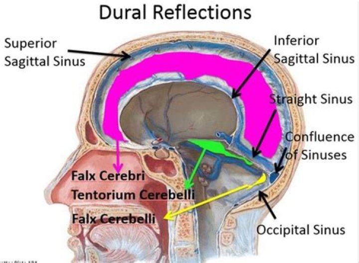
Dural reflections refer to places where two face-to-face meningeal layers descend into the cranial cavity to form the septa that compartmentalize the brain. The two main dural reflections are the falx cerebri and the tentorium cerebelli.
Which dural reflections contain sinuses?
- Two horizontal reflections.
- Superior sagittal sinus: lies at the superior attached border of falx cerebri.
- Inferior sagittal sinus: lies at the inferior free border of falx cerebri.
- Straight sinus: lies at the line of attachment of falx cerebri and tentorium cerebelli.
What is the function of dural folds?
The meningeal layer of the dura mater creates several dural folds that divide the cranial cavity into freely communicating spaces. The function of the dural folds is to limit the rotational displacement of the brain. The folds include the following: The falx cerebri is a meningeal projection of dura in the brain.
What is the dural of the brain?
The dura mater often gets referred to as merely the dura. It is one of the layers of connective tissue that make up the meninges of the brain (pia, arachnoid, and dura, from inside to outside). It is the outermost layer of the three meninges that surround and protect the brain and spinal cord.What is the purpose of dura mater?
The dura mater is a sac that envelops the arachnoid and has been modified to serve several functions. The dura mater surrounds and supports the large venous channels (dural sinuses) carrying blood from the brain toward the heart. The dura mater is partitioned into several septa, which support the brain.
Is the dura mater pain sensitive?
In this observational study, we confirmed that dura of the skull base and dura of the falx cerebri are sensitive to pain and that their mechanical stimulation induced pain mainly referred in the sensory territories of the V1 and V3 divisions of the trigeminal nerve.
What does it mean when the dural venous sinuses are patent?
On conventional MRI sequences, patent dural sinuses are often seen as a flow void. This is particularly well seen when the imaging plane is orthogonal to the blood flow direction (e.g., coronal images are best for visualization of the superior sagittal, transverse, and sigmoid sinuses).
Where is your dura?
Dura mater is a thick membrane made of dense irregular connective tissue that surrounds the brain and spinal cord. It is the outermost of the three layers of membrane called the meninges that protect the central nervous system. The other two meningeal layers are the arachnoid mater and the pia mater.What is dural?
dural. / (ˈdjʊərəl) / adjective. relating to or affecting the dura mater.
What is the dura in the neck?It is the container for the cerebrospinal fluid that flows from the brain into the spinal canal. The dura is present in the neck from the C1 to C3 cervical vertebrae. When these cervical vertebrae are unstable and wandering, they cause tension on the muscles, which are in fact trying to hold the vertebrae in place.
Article first time published onDo the dural venous sinuses lack valves?
Unlike other blood vessels, dural venous sinuses lack valves and other vessel associated layers. Large endothelium-lined venous channels situated between the two layers of DURA MATER, the endosteal and the meningeal layers. They are devoid of valves and are parts of the venous system of dura mater.
How does the dura mater protect the brain?
The dura provides the brain and spinal cord with an extra protective layer, helps to keep the CNS from being jostled around by fastening it to the skull or vertebral column, and supplies a complex system of veinous drainage through which blood can leave the brain.
What is a dural graft?
dura. graft – material, or tissue, surgically implanted into a body. part to replace or repair a defect.
What are the two main functions of the CSF?
CSF provides hydromechanical protection of the neuroaxis through two mechanisms. First, CSF acts as a shock absorber, cushioning the brain against the skull. Second, CSF allows the brain and spinal cord to become buoyant, reducing the effective weight of the brain from its normal 1,500 grams to a much lesser 50 grams.
Where is CSF produced?
CSF is produced mainly by the choroid plexus epithelium and ependymal cells of the ventricles and flows into interconnecting chambers; namely, the cisterns and the subarachnoid spaces.
What is the function of the arachnoid?
Arachnoid mater: Connected to the dura mater on the side closest to the CNS, this middle layer includes a network of fibers and collagen that are part of the suspension system that helps protect the brain and spinal cord from sudden impact.
Where do the dural venous sinuses drain?
The dural venous sinuses lie between the periosteal and meningeal layers of the dura mater. They are best thought of as collecting pools of blood, which drain the central nervous system, the face, and the scalp. All the dural venous sinuses ultimately drain into the internal jugular vein.
How do the dural venous sinuses differ from veins?
The walls of the dural venous sinuses are composed of dura mater lined with endothelium, a specialized layer of flattened cells found in blood vessels. They differ from other blood vessels in that they lack a full set of vessel layers (e.g. tunica media) characteristic of arteries and veins.
What is a venous sinus in the brain?
venous sinus, in human anatomy, any of the channels of a branching complex sinus network that lies between layers of the dura mater, the outermost covering of the brain, and functions to collect oxygen-depleted blood. Unlike veins, these sinuses possess no muscular coat.
Why the brain is painless?
The brain itself does not feel pain because there are no nociceptors located in brain tissue itself. This feature explains why neurosurgeons can operate on brain tissue without causing a patient discomfort, and, in some cases, can even perform surgery while the patient is awake.
What are dural signs?
Dural signs and symptoms are those that are related to the increase of dural irritation: traction exerted from a distance (straight leg raising and neck flexion) pulls on the inflamed dura or, via the dural ligaments, on the posterior longitudinal ligament or outer annular rim.
Is dura mater attached to skull?
The dura mater is firmly attached to the rim of the foramen magnum and its fibres blend with the periosteum within the skull. In the spinal canal it is not attached to the vertebral arches, because of the presence of protective fat tissue in between.
Is dural a word?
Yes, dural is in the scrabble dictionary.
Why is it called arachnoid mater?
The arachnoid mater, named for its spiderweb-like appearance, is a thin, transparent membrane surrounding the spinal cord like a loosely fitting sac.
What is the subarachnoid space?
The subarachnoid space consists of the cerebrospinal fluid (CSF), major blood vessels, and cisterns. The cisterns are enlarged pockets of CSF created due to the separation of the arachnoid mater from the pia mater based on the anatomy of the brain and spinal cord surface.
What are the 3 layers of the brain?
Three layers of membranes known as meninges protect the brain and spinal cord. The delicate inner layer is the pia mater. The middle layer is the arachnoid, a web-like structure filled with fluid that cushions the brain. The tough outer layer is called the dura mater.
Does the dura have nerves?
The cerebral dura mater is richly innervated by afferent nerve fibers, most of which originate in the ipsilateral trigeminal ganglion, and by sympathetic fibers predominantly arising from the ipsilateral superior cervical ganglion 3,4.
What happens if the dura mater tears?
Cerebrospinal fluid (CSF) leakage following dural tears can pose potentially serious problems such as CSF fistula formation, pseudomeningocele, meningitis, arachnoiditis and epidural abscess[1,3,10,12,15].
Where is the dura in the spine?
The dura is a thin layer of tissue that covers and protects the spinal cord. It lies in between the spine (the bone) and the spinal cord (nerve tissue).
Which is the largest among the cranial venous sinuses?
Superior sagittal sinus—this is the largest dural venous sinus. It runs in a sagittal plane, from the anterior falx cerebri to its point of termination at the occipital protuberance.
What do the dural venous sinuses contain?
The Dural Venous Sinuses. The dural venous sinuses are spaces between the endosteal and meningeal layers of the dura. They contain venous blood that originates for the most part from the brain or cranial cavity. The sinuses contain an endothelial lining that is continuous into the veins that are connected to them.