What is a cryostat machine

Robert Spencer
Published Mar 16, 2026
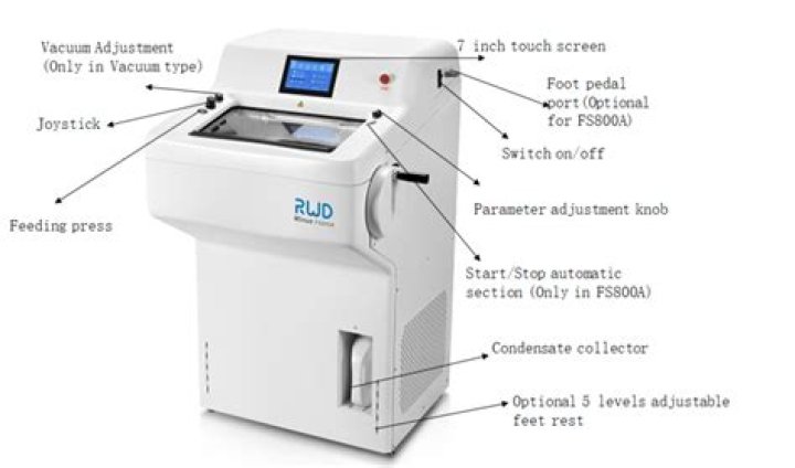
A cryostat is a microtome machine for cutting tissue at low temperatures (typically around −15 to −30°C) (Figure 55).
What is the difference between a cryostat and microtome?
What is a Cryostat? Similar to a standard microtome, a cryostat functions to obtain thin (1-10 mm in thickness) sections from a piece of tissue, but while a standard microtome carries the operation at room temperature, the cryostat enables the operator to section the tissue at low temperature (–20 to –30 C).
How much is a cryostat?
Cryostats generally cost in the region of USD$8,000 to $12,000.
Which gas is in cryostat?
Closed cycle helium cryostats use high-pressure helium gas to produce cooling.What are the advantages of cryostat?
The advantage of cryostat-cut frozen section is the preservation of cell density and tissue architecture, which is most important in the diagnosis of neurosurgical materials, especially in case of diffusely infiltrating glioma.
How do cryostats work?
A cryostat is a machine that maintains the cryogenic temperature of samples or devices placed inside it. … Cryostats use a number of refrigeration methods to achieve low temperatures, such as a helium bath with liquid helium or liquid nitrogen. Other cryogenic fluids are argon, oxygen, and nitrogen.
What are histological features?
Histology, also known as microscopic anatomy or microanatomy, is the branch of biology which studies the microscopic anatomy of biological tissues. Histology is the microscopic counterpart to gross anatomy, which looks at larger structures visible without a microscope.
How do you maintain a cryostat?
CRYOSTAT MAINTENANCE25 Remove the microtomy debris from the cryostat chamber and place into a fixative, disinfectant or wet bag for autoclaving. Clean the knife and all exposed surfaces and instruments with 70% ethanol. Wear protective clothing as for routine maintenance. Turn off the power and unplug the cryostat.When was cryostat invented?
The invention of the first performing cryostats is generally attributed to Sir James Dewar (Fig. 1), and hence cryostats containing cryogenic fluids are nowadays also called dewars. In 1897 Dewar used silver-plated double-walled glass containers to collect the first liquefied hydrogen.
How do I choose a cryostat?If you’re seeking a new cryostat for your lab, it’s essential to select a reliable device with adequate temperature control and ultra-thin sectioning capabilities. The ideal cryostat has all the features you need to offer fast results and create an efficient workflow.
Article first time published onHow do you freeze tissue for cryostat sectioning?
Rapidly wrap all samples in pre-cooled, labeled foil, and place in a pre-cooled plastic bag, in a freezer box. Store at -80°C. temperature for approximately 30 minutes. the tissues have been freshly frozen or pre-fixed with subsequent cryoprotection.
What is temperature of cryostat?
A cryostat is a microtome machine for cutting tissue at low temperatures (typically around −15 to −30°C) (Figure 55).
What are the types of Microtomes?
- hand microtome.
- rotational microtome.
- sledge microtome.
- cryostat (freezing microtome)
- ultramicrotome.
Why cryostat sectioning is used to analyze the tissue during surgery?
Cryostat Sectioning Look at the clinical information available as it can help achieve the possible differential diagnoses. Observe the gross appearance of the tissue in terms of color, consistency, texture, and presence of sutures used to mark the anatomical position of the sample.
What is histological technique?
In order to study tissues with a microscope they must be preserved (fixed) and cut into sections thin enough to be translucent. Fundamentally it consists of a chemical or physical method of killing the tissue and yet retaining characteristic peculiarities of shape and structure. …
What is histological anatomy?
Definition of histology 1 : a branch of anatomy that deals with the minute structure of animal and plant tissues as discernible with the microscope. 2 : tissue structure or organization. Other Words from histology Example Sentences Learn More About histology.
Is histopathology and histology same?
The National Cancer Institute defines histopathology as “the study of diseased cells and tissues using a microscope.”1 Histology is the study of tissues, and pathology is the study of disease. So taken together, histopathology literally means the study of tissues as relates to disease.
What is closed cycle cryostat?
Advanced Research Systems closed cycle cryocoolers, also known as dry cryostats, allow a system to be cooled down to liquid helium temperatures without the use of cryogens. The only utilities required are electricity and cooling water for the compressor. These cryostats are sometimes referred to as ‘helium-free.
What is a Cryomicrotome?
Cryomicrotome is a sectioning instrument that allows for the cutting of extremely thin sections upto 20 micron of frozen tissue or scaffold for microscopy and other applications.
What is a vibrating microtome?
Vibrating microtomes (Vibratomes) by Leica Biosystems help you to accurately cut tissue under physiologial conditions without freezing or embedding. These precise instruments maintain cell morphology, enzyme activity, as well as cell viability of the tissue.
How does a cryocooler work?
In most cases cryocoolers use a cryogenic fluid as the working substance and employ moving parts to cycle the fluid around a thermodynamic cycle. … The returning low-pressure fluid passes through the heat exchanger to precool the high-pressure fluid before entering the compressor intake. The cycle is then repeated.
Who invented the cryostat?
Sir James Dewar, Scottish Physicist, and Chemist are credited for the use of the very first cryostats in the 19th century.
What is the principle of automatic tissue processor?
Automated tissue processors are of two main types: tissue-transfer machines and fluid-transfer machines. The tissue processor finds applications in histopathology laboratories to automatically prepare tissue samples for laboratory testing, by fixing, dehydrating, clearing, and infiltrating them with paraffin.
What is cryostat in histopathology?
Cryostats are used in medicine to cut histological slides. They are usually used in a process called frozen section histology (see Frozen section procedure). The cryostat is essentially an ultrafine “deli-slicer”, called a microtome, placed in a freezer.
What is frozen section PPT?
What is frozen section? The frozen section procedure is a pathological laboratory procedure to perform rapid microscopic analysis of a specimen. The technical name for this procedure is Cryo section.
How long does it take for brains to sink in sucrose?
Your brain sections cracked maybe a result of not being dehydrated properly in the sucrose solution. For adult mouse brains, it takes at least 3 days for dehydration to complete – i.e. the brain sinks to the bottom of the tube in 30% sucrose.
Can you snap freeze with dry ice?
Snap freezing, or flash freezing, is the process by which samples are lowered to temperatures below -70°C very rapidly using dry ice or liquid nitrogen. Snap freezing achieves the same endpoint as slow rate-controlled freezing, but at approximate rate of -10-1000°C/min, compared to -1°C/min.
How do you get rid of October?
OCT can just be gently rinsed away. If section thickness is important to you, be sure to limit the exposure to alcohols and air – both can cause a tremendous amount of shrinkage.
What is the optimum cryostat temperature for uterine tissue?
Optimum temperature —15 to —25°C: Muscle, connective tissue, uterus, cervix, pancreas, skin without fat, prostate, ovary, gut, nonfatty breast tissue.
What is trimming of tissue block?
Trimming is cutting a fixed tissue or organ to create a flat surface with correct orientation. You can do this yourself with training and a few basic supplies (cutting board, forceps, single edge razor blade) or our histologists can do this for you. Trimming should be done after, not prior to, fixation.
How do microtomes work?
The vibrating microtome operates by cutting using a vibrating blade, allowing the resultant cut to be made with less pressure than would be required for a stationary blade. The vibrating microtome is usually used for difficult biological samples.