What does VQ mismatch mean

Emily Dawson
Published Mar 15, 2026
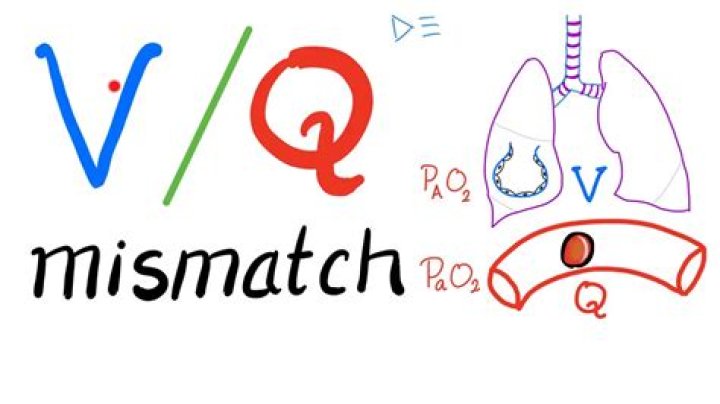
What a V/Q mismatch means. A V/Q mismatch happens when part of your lung receives oxygen without blood flow or blood flow without oxygen. This happens if you have an obstructed airway, such as when you’re choking, or if you have an obstructed blood vessel, such as a blood clot in your lung.
What is meant by VQ mismatch?
What a V/Q mismatch means. A V/Q mismatch happens when part of your lung receives oxygen without blood flow or blood flow without oxygen. This happens if you have an obstructed airway, such as when you’re choking, or if you have an obstructed blood vessel, such as a blood clot in your lung.
What are the types of VQ mismatch?
V/Q mismatch is common and often effects our patient’s ventilation and oxygenation. There are 2 types of mismatch: dead space and shunt. Shunt is perfusion of poorly ventilated alveoli. Physiologic dead space is ventilation of poor perfused alveoli.
What conditions causes VQ mismatch?
Some common causes of hypoxemia due to V/Q mismatch include asthma, COPD, bronchiectasis, cystic fibrosis, interstitial lung diseases (ILDs), and pulmonary hypertension.Is VQ mismatch normal?
A normal V/Q ratio is around 0.80. Roughly four liters of oxygen and five liters of blood pass through the lungs per minute. A ratio above or below 0.80 is considered abnormal. 3 Higher-than-normal results indicate reduced perfusion; lower-than-normal results indicate reduced ventilation.
Is ARDS shunt or dead space?
Acute respiratory distress syndrome (ARDS) is characterized by severe impairment of gas exchange. Hypoxemia is mainly due to intrapulmonary shunt, whereas increased alveolar dead space explains the alteration of CO2 clearance.
Is PE shunt or dead space?
Pulmonary embolism (PE) is an example of increased dead space resulted in decreasing perfusion relative to ventilation. Shunt and dead space are two conditions of lungs, resulting in impaired gas exchange. Moreover, they are examples of the ventilation-perfusion (V/Q) mismatch.
How is a pulmonary shunt treated?
- Treatment.
- Oxygen Therapy.
- Mechanical Ventilation.
- Positive End-Expiratory Pressure.
- Body Positioning.
- Nitric Oxide.
- Long-Term Oxygen Therapy.
- Exercises.
Is atelectasis serious?
Large areas of atelectasis may be life threatening, often in a baby or small child, or in someone who has another lung disease or illness. The collapsed lung usually reinflates slowly if the airway blockage has been removed. Scarring or damage may remain. The outlook depends on the underlying disease.
How do you improve gas exchange in the lungs?Improvements in gas exchange occur via several mechanisms: alterations in the distribution of alveolar ventilation, redistribution of blood flow, improved matching of local ventilation and perfusion, and reduction in regions of low ventilation/perfusion ratios.
Article first time published onIs PE a shunt?
In acute PE, intracardiac shunting usually occurs through a patent foramen ovale; right atrial pressure exceeds left atrial pressure, even if both pressures are normal.
Is atelectasis a shunt or V Q mismatch?
The major cause of this derangement is shunt, an effect of prompt atelectasis formation in dependent lung regions. An additional cause is ventilation/perfusion (V/Q) mismatch, possibly produced by intermittent airway closure.
What is the difference between shunt and VQ mismatch?
A , VQ mismatch occurs with regional differences in the optimal alveolar-capillary interface as gas exchange occurs unimpeded (wide arrow) in some areas and restricted (narrow arrow) or prohibited (X) in others. … B , Shunt occurs when blood fl ow does not participate in gas exchange, such as is observed with ARDS.
Which situation will happen when you have emphysema?
In emphysema, the inner walls of the lungs’ air sacs (alveoli) are damaged, causing them to eventually rupture. This creates one larger air space instead of many small ones and reduces the surface area available for gas exchange. Emphysema is a lung condition that causes shortness of breath.
How does COPD affect ventilation?
In COPD, the airways of the lungs (bronchial tubes) become inflamed and narrowed. They tend to collapse when you breathe out and can become clogged with mucus. This reduces airflow through the bronchial tubes, a condition called airway obstruction, making it difficult to move air in and out of the lungs.
How do VQ scans detect pulmonary embolism?
It uses special x ray scanners outside of your body to create pictures of air and blood flow patterns in your lungs. This test can help diagnose or rule out a pulmonary embolism, or a blood clot in your lung. A VQ scan also can detect regional differences in lung blood flow and air distribution.
Does oxygen help pulmonary embolism?
Treatment goals for pulmonary embolism are to improve oxygenation and cardiac output. Administer supplemental oxygen via nasal cannula or non-rebreather mask to maintain SPO2 above 94 percent. Be aware that reduced blood flow to the lungs may prevent improvement of hypoxia from oxygen administration.
Does a shunt respond to oxygen?
True shunt is refractory to oxygen therapy. This results in what is termed “refractory hypoxemia”. Because refractory hypoxemia does not respond to oxygen therapy, other means should be sought to improve arterial oxygenation.
What is a relative shunt?
We define a right-to-left relative shunt in the pulmonary circulation as one in which mixed venous blood flows through the capillary beds of alveoli whose ventilation is inadequate to arterialize the blood. The excess blood flow through these regions!
Why does shunt not respond to oxygen?
Because shunt represents areas where gas exchange does not occur, 100% inspired oxygen is unable to overcome the hypoxia caused by shunting. … Although ventilation at that area is unaffected, blood will not be able to flow through that capillary; therefore, at that zone there will be no gas exchange.
Does shunt cause hypercapnia?
Shunt as a cause of hypoxemia is observed primarily in pneumonia, atelectasis, and severe pulmonary edema of either cardiac or noncardiac origin. Hypercapnia generally does not develop unless the shunt is excessive (> 60%).
How do you treat dead space in the lungs?
Mechanical Ventilation: Tubing from the ventilator increases dead space volume by adding volume to the effective space, not participating in gas exchange. PEEP: Excessive PEEP can over-distend alveoli and result in lung barotrauma, increasing the dead space volume.
What are the 3 types of atelectasis?
There are three major types of atelectasis: adhesive, compressive, and obstructive.
What is the treatment for Subsegmental atelectasis?
Atelectasis treatments include: Bronchoscopy to clear blockages like mucus. Medicine that you breathe in through an inhaler. Physiotherapy such as tapping on your chest to break up mucus, lying on one side or with your head lower than your chest to drain mucus, and exercises to help you breathe better.
How do you fix atelectasis?
Atelectasis treatment can include breathing or coughing exercises, inhaled medicines, breathing devices, or surgery. Atelectasis usually gets better with time or treatment. However, if it is undiagnosed or untreated, serious complications can occur, including fluid buildup, pneumonia, and respiratory failure.
What is right-to-left shunt in lungs?
A shunt is an abnormal communication between the right and left sides of the heart or between the systemic and pulmonary vessels, allowing blood to flow directly from one circulatory system to the other. A right-to-left shunt allows deoxygenated systemic venous blood to bypass the lungs and return to the body.
What is shunt in the lungs?
“Shunt” means decreased ratios and includes perfused alveoli without ventilation; very poorly ventilated alveoli with normal, increased, or slightly decreased perfusion; and ventilated alveoli with markedly increased perfusion.
Why Does Dead Space correct with oxygen?
Dead space is the volume of air that is inhaled that does not take part in the gas exchange, because it either remains in the conducting airways or reaches alveoli that are not perfused or poorly perfused. It means that not all the air in each breath is available for the exchange of oxygen and carbon dioxide.
What is the best position for a patient in respiratory distress?
Prone positioning is widely used to improve oxygenation of patients with acute respiratory distress syndrome (ARDS).
What are the 4 stages of COPD?
- Stage 1: Mild COPD. …
- Stage 2: Moderate COPD. …
- Stage 3: Severe COPD. …
- Stage 4: Very Severe COPD. …
- Early Detection and Smoking Cessation. …
- Get the treatment you need to slow the progression of COPD.
What causes poor gas exchange in lungs?
By far the commonest cause of impaired gas exchange in patients with lung disease is ventilation-perfusion inequality. This is a complicated topic and much can be learned from computer models. Ventilation-perfusion inequality always causes hypoxemia, that is, an abnormally low PO2 in arterial blood.