What does an ABR test show

Mia Kelly
Published Apr 09, 2026
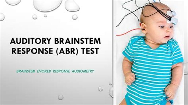
The auditory brainstem response (ABR) test tells us how the inner ear, called the cochlea, and the brain pathways for hearing are working. You may also hear it called an auditory evoked potential (AEP). The test is used with children or others who cannot complete a typical hearing screening.
How long does an ABR test Take for adults?
An ABR test usually takes 1–2 hours, but the appointment may last about 3 hours.
Can ABR detect tinnitus?
This is one of many tests that may be recommended for the evaluation of dizziness, balance problems, hearing loss, ear pressure and/or tinnitus (ear noise). The ABR determines how well the auditory nerve is transmitting sound (in the form of electrical impulses) from the inner ear to the brainstem.
What does an abnormal ABR mean?
An abnormal ABR may be a consistent finding with high frequency hearing loss or profound deafness, it may also be an indication that further testing is required. However, only the referring physician will be able to determine if further tests or procedures are required. Caring, Competence, and Credentials TM.How is an ABR test performed?
How Is an ABR Done? An audiologist places small earphones in the child’s ears and soft electrodes (small sensor stickers) near the ears and on the forehead. Clicking sounds and tones go through the earphones, and electrodes measure how the hearing nerves and brain respond to the sounds.
Is ABR test safe?
The ABR test is safe and does not hurt. The ABR test can be completed only if the child is sleeping or lying perfectly still, relaxed and with his or her eyes closed. If your child is younger than 6 months of age, the ABR test usually can be done while he or she naps.
Who performs an ABR test?
This test is painless and noninvasive. The audiologist attaches adhesive recording electrodes on the forehead and ears, and captures and analyzes recordings of electric potentials generated by the auditory neural pathway — the network of nerves that move from the ears to the brain.
What happens during a sedated ABR?
During a Sedated ABR test Once the child is asleep, the audiologist gently cleans the skin on his / her forehead and behind each ear and small sensors are placed on those areas. Sound is sent through the earphones and the sensors measure the brain’s response to those sounds.Can ABR detect conductive hearing loss?
The results showed that the hearing loss (conductive hearing loss of sersorineural hearing loss) can be detected by ABR with the stimulation of pure tone in the range of 2000 to 4000 Hz.
What is the function of auditory brainstem?This is most commonly due to a missing or very small hearing nerve or severely abnormal inner ear (cochlea). The auditory brainstem implant directly stimulates the hearing pathways in the brainstem, bypassing the inner ear and hearing nerve.
Article first time published onHow much does a sedated ABR cost?
Assuming a 90-minute time slot, we estimate an average cost of sedation for ABR at $2,043 per occurrence. Failure to sedate sufficiently while accomplishing the procedure through restraint and failing to complete the task through inadequate sedation will lead to additional costs in the post-anesthetic care unit (PACU).
Can ABR results change?
The time between the measurements was on average 5 months (0 to 31 months). Conclusion: Hearing threshold changes are often seen in repeated ABR measurements. Therefore multiple measurements are necessary when ABR yields abnormal. Hearing threshold changes should be taken into account for hearing aid provision.
What organs are in the auditory system?
The auditory system processes how we hear and understand sounds within the environment. It is made up of both peripheral structures (e.g., outer, middle, and inner ear) and brain regions (cochlear nuclei, superior olivary nuclei, lateral lemniscus, inferior colliculus, medial geniculate nuclei, and auditory cortex).
What causes hearing loss due to nerve damage?
The nerves then carry these signals to the brain. Sensorineural hearing loss (SNHL) is caused by damage to these special cells, or to the nerve fibers in the inner ear. Sometimes, the hearing loss is caused by damage to the nerve that carries the signals to the brain.
How long does a BAER test take?
Each ear is tested individually, and the test usually is complete in 10-15 minutes. Sedation or anesthesia are usually not necessary unless the dog becomes extremely agitated, which can usually be avoided with patient and gentle handling.
Why Bera test is done?
Brainstem-evoked response audiometry (BERA) is a simple, noninvasive, objective test for early identification of hearing impairment in children and neonates. It can be used as a screening test and is useful in newborns, infants, and other difficult-to-test patients.
How accurate are ABR tests?
ABR accuracy is excellent for detecting average sensorineural hearing loss at 2 and 4 kHz in excess of 30 dB, and the overall results for a wide range of hearing loss and ABR abnormality criteria can be conveniently summarized in terms of relative operating characteristics (ROCs).
What is OAE in audiology?
The OAE test is used to find out how well your inner ear, or cochlea, works. It measures otoacoustic emissions, or OAEs. These are sounds given off by the inner ear when responding to a sound.
What is the auditory pathway to the brain?
Auditory messages are conveyed to the brain via two types of pathway: the primary auditory pathway which exclusively carries messages from the cochlea, and the non-primary pathway (also called the reticular sensory pathway) which carries all types of sensory messages.
Do auditory nerves cross over?
Once they leave the cochlear nucleus, most of the axons of the cochlear nucleus cells cross over to the opposite side (contralateral side) of the brain (Figure 27 ). This means that most of the auditory information processed by each half of the brain comes from the ear on the other side of the head.
Where is the auditory nerve located?
The cochlear nerve, also known as the acoustic or auditory nerve, is the cranial nerve responsible for hearing. It travels from the inner ear to the brainstem and out through a bone located on the side of the skull called the temporal bone.