What do Osborn waves mean

Christopher Lucas
Published Feb 16, 2026
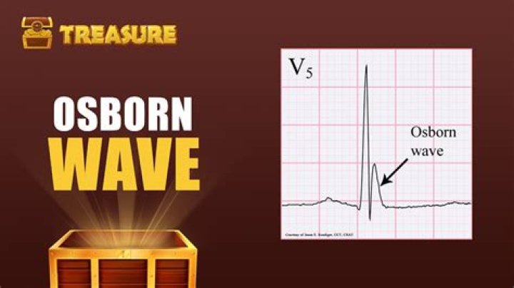
Osborn Wave (J Wave) Overview The Osborn wave (J wave) is a positive deflection at the J point (negative in aVR and V1). It is usually most prominent in the precordial leads. Eponymously associated with John Jay Osborn (1917-2014) following his 1953 ‘current of injury’ description in hypothermic dogs.
What is associated with Osborne or J waves on the ECG?
Osborn wave (J wave). These waves occur due to hypothermia, hypercalcemia, early repolarization and Brugada syndrome.
What does J point elevation indicate?
The term J-point elevation represents a family of ECG findings. It has been described in several metabolic disorders most notably hypothermia (abnormally low body temperature). Subtle nuances in its pattern may point to other conditions, the most common of which is termed ‘early repolarization’.
Are J Waves bad?
The J wave is a positive deflection in the electrocardiogram (ECG) that occurs at the junction between the QRS complex and the ST segment, also known as the J point.When do you see Osborn waves?
First described in 1938, this electrocardiographic feature is also known as the Osborn wave or hypothermic hump. It is seen at the junction of the QRS and ST segments and may appear at temperatures below 32°C. It is most often seen in leads II and V6, but in more severe hypothermia may be seen in V3 or V4.
What is J point depression?
Elevation or depression of the J point is seen with the various causes of ST segment abnormality. It may be elevated as a result of injury currents during acute myocardial ischemia and pericarditis, as well as in various other patterns of both normal and abnormal ECGs.
What is early repolarization ECG?
Early repolarization pattern (ERP) is a common ECG variant, characterized by J point elevation manifested either as terminal QRS slurring (the transition from the QRS segment to the ST segment) or notching (a positive deflection inscribed on terminal QRS complex) associated with concave upward ST-segment elevation and …
What is STJ in ECG?
Key definitions. STEMI (ST elevation myocardial ischemia/infarction) STJ level (ST level at J point, QRS end) STEMI imposter (non-ischemic cause of ST elevation)What is Brugada syndrome?
Brugada (brew-GAH-dah) syndrome is a rare, but potentially life-threatening heart rhythm disorder that is sometimes inherited. People with Brugada syndrome have an increased risk of having irregular heart rhythms beginning in the lower chambers of the heart (ventricles).
What does J point notching mean?Early repolarization syndrome ER is characterized by prominent J-point on ECG with notching/slurring of distal part of R wave which more or less appears as pseudo delta wave. J notches are known for long time and are actually present in 2–10% of general population.
Article first time published onWhere is the J point on the ECG?
Introduction. The J-point on the electrocardiographic waveform is historically defined as the junction between the end of the QRS complex and the beginning of the ST-segment.
What are the symptoms of hypothermia?
- Shivering.
- Exhaustion or feeling very tired.
- Confusion.
- Fumbling hands.
- Memory loss.
- Slurred speech.
- Drowsiness.
What are the ECG abnormalities in hypothermia?
The ECG findings of hypothermia include: An “Osborne wave” characterized by a notch in the downward portion of the R wave in the QRS complex. Low voltage. Bradycardia: This can be sinus bradycardia, junctional bradycardia, atrial fibrillation with a slow ventricular response or higher grade AV blocks.
Is early repolarization an arrhythmia?
Early repolarization syndrome (ERS), demonstrated as J-point elevation on an electrocardiograph, was formerly thought to be a benign entity, but the recent studies have demonstrated that it can be linked to a considerable risk of life – threatening arrhythmias and sudden cardiac death (SCD).
What are the symptoms of early repolarization?
Life-threatening arrhythmias are often the first, unexpected clinical manifestation of early repolarization syndrome. An increase of J wave/ST segment amplitude has been described before the onset of ventricular fibrillation in early repolarization patients.
Does benign early repolarization go away?
Temporal Stability of BER The ST elevation may gradually disappear over time as the patient ages: up to 30% of patient with BER will have resolution of ST elevation on ECGs taken many years later.
What does inverted T wave mean on ECG?
Inverted T waves. Ischemia: Myocardial ischemia is a common cause of inverted T waves. Inverted T waves are less specific than ST segment depression for ischemia, and do not in and of themselves convey a poor prognosis (as compared to patients with an acute coronary syndrome and ST segment depression).
Are you born with Brugada syndrome?
Brugada syndrome is an unusual genetic disorder of the heart’s electrical system. Although people are born with it, they usually do not know they have it until they reach their 30s or 40s. The only symptoms of Brugada syndrome are passing out (called syncope), or heart palpitations, or sudden cardiac death.
Can you live a full life with Brugada syndrome?
It can do, although many people with Brugada syndrome can lead an entirely normal life.
Is Brugada an arrhythmia?
Brugada syndrome is a condition that causes a disruption of the heart’s normal rhythm . Specifically, this disorder can lead to irregular heartbeats in the heart’s lower chambers (ventricles), which is an abnormality called ventricular arrhythmia.
What does a prolonged PR interval indicate?
A prolonged PR interval represents a delay in the time it takes for the signal to move across the atria at the top of the heart, which receive blood flowing in from the veins, into the ventricles at the bottom of the heart, which pump blood out into the arteries.
Can ECG detect heart blockage?
However, it does not show whether you have asymptomatic blockages in your heart arteries or predict your risk of a future heart attack. The resting ECG is different from a stress or exercise ECG or cardiac imaging test.
What does a spike on an EKG mean?
For example, spikes that are too close together are a sign of a rapid heartbeat or tachycardia. Each heartbeat will be made up of several spikes in activity. The first P wave shows when the atria are contracting. The second and biggest spike, known as the QRS complex, occurs when your ventricles contract.
Why do athletes get early repolarization?
Early repolarization (ERP) is a common finding in young, healthy, competitive athletes and appears to be a direct result of exercise training. Both ERP and an inferior subtype (originally thought to increase risk of sudden death) increase in prevalence after intense physical training.
Which ECG finding is characterized by elevation of the J point?
Early repolarization – UpToDate. The term early repolarization (ER), also known as “J-waves” or “J-point elevation,” has long been used to characterize a QRS-T variant on the electrocardiogram (ECG).
Which of the following is a historical figure for whom the J point wave was named after in 1953?
History. The prominent J deflection attributed to hypothermia was first reported in 1938 by Tomaszewski. These waves were then definitively described in 1953 by John J. Osborn (1917–2014) and were named in his honor.
What happens during the T wave?
The T wave represents ventricular repolarization. Generally, the T wave exhibits a positive deflection. The reason for this is that the last cells to depolarize in the ventricles are the first to repolarize.
How long is a tele strip?
Each small square is 1 mm in length and represents 0.04 seconds. Each larger square is 5 mm in length and represents 0.2 seconds.
What temperature is too low for a person?
Body temperature below 95°F (35°C) is considered abnormally low, and the condition is known as hypothermia. This happens when your body loses heat faster than it can produce heat. Hypothermia is a medical emergency, which if left untreated can lead to brain damage and cardiac failure.
What happens to the brain during hypothermia?
Hypothermia progressively depresses the CNS, decreasing CNS metabolism in a linear fashion as the core temperature drops. At core temperatures less than 33°C, brain electrical activity becomes abnormal; between 19°C and 20°C, an electroencephalogram (EEG) may appear consistent with brain death.
What are the 5 stages of hypothermia?
- HT I: Mild Hypothermia, 95-89.6 degrees. Normal or nearly normal consciousness, shivering.
- HT II: Moderate Hypothermia, 89.6-82.4 degrees. …
- HT III: Severe Hypothermia, 82.4-75.2 degrees. …
- HT IV: Apparent Death, 75.2-59 degrees.
- HT V: Death from irreversible hypothermia.