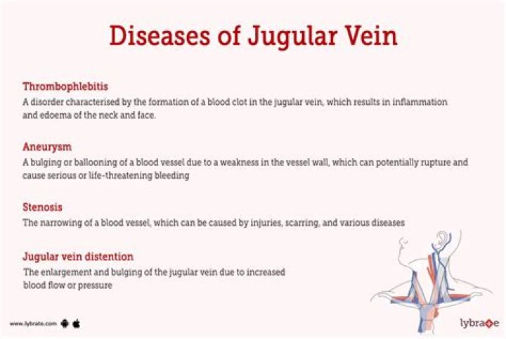
What causes increased JVD

Rachel Hickman
Published Feb 18, 2026
JVD is often caused by life-threatening conditions such as pulmonary embolism, tension pneumothorax, car- diac tamponade, and heart failure,1 and is a classic and crucial finding in the evaluation of all patients presenting with shock.
What does jugular venous pressure indicate?
Elevated jugular venous pressure is a manifestation of abnormal right heart dynamics, mostly commonly reflecting elevated pulmonary capillary wedge pressure from left heart failure. 12. This usually implies fluid overload, indicating the need for diuresis.
Why do we assess for JVD?
Its most common use is to estimate the right atrial pressure, especially in patients with heart failure, and also to measure the response to diuretic therapy. It is also helpful in the evaluation of conditions such as superior vena cava obstruction, tricuspid valve disease, and pericardial disease.
What do distended neck veins indicate?
In patients with acute inferior-wall MI with right ventricular involvement, distention of neck veins is commonly described as a sign of failure of the right ventricle. Impaired right ventricular function also leads to systemic venous hypertension, edema, and hepatomegaly.How serious is JVD?
JVD can be the sign of a severe condition, including heart failure, so it is vital that a person is seen by a medical professional as soon as possible. While heart failure can happen to anyone, risk factors for heart failure include: high blood pressure.
What causes JVD in CHF?
Common causes of jugular vein distention Congestive heart failure (deterioration of the heart’s ability to pump blood) Constrictive pericarditis (infection or inflammation of the lining that surrounds the heart that decreases the lining’s flexibility) Hypervolemia (increased blood volume)
How do you evaluate JVD?
To properly evaluate jugular venous distension, the patient must be placed at a 45-degree angle, or slightly less. Visualization of the jugular veins is best done at an oblique angle, so sit beside the patient and elevate the head of the cot into a semi-Fowler’s position.
What is considered elevated JVP?
JVP is > 9 cm above the right atrium (> 4 cm above the sternal angle)What is the normal venous pressure?
A normal central venous pressure reading is between 8 to 12 mmHg. This value is altered by volume status and/or venous compliance.
Is an EJ a central line?Central venous cannulation via the external jugular vein (EJV) is a recognized technique [1-3]. It is associated with minimal complications but with a relatively frequent failure rate compared with the cannulation of the internal jugular or subclavian veins (SCV) [1,3,4].
Article first time published onHow do nurses test for JVD?
Measure the height of the bulge or distention of the jugular vein. Ask the patient to lie down on the exam table with the head at 45 degrees. Ask him/her to turn his/her head to the side to measure the central venous pressure (CVP). The physician will measure the height of the bulge of JVD to indicate CVP.
How do you know if your JVP is high?
It has been taught that the best method for evaluating the JVP is to position the patient supine in bed, elevate the patient’s head to approximately 30–45 degrees, and measure or estimate the vertical height of the meniscus of the right internal or external jugular vein above the sternal angle (angle of Louis) which is …
Does a pneumothorax cause JVD?
Thus, a tension pneumothorax creates not only a respiratory compromise but also a cardiovascular compromise. Tension pneumothorax presents with respiratory distress, jugular venous distention (JVD), diminished breath sounds, tachycardia and narrow pulse pressure.
How would you describe JVD on a physical exam?
Venous wave is bifid, flicking like a snake’s tongue. It rises when you lower the head of the bed and sinks when you raise the head of the bed. It changes with respiration, sinking into the chest with inspiration. It is not palpable.
How is CHF different than COPD?
Outlook. COPD and CHF are serious conditions that affect your breathing and can affect your activity in life. Although both have similar symptoms and risk factors, COPD affects your lungs and CHF affects your heart. Different medications are used to treat each condition.
What does a high map mean?
A high MAP is anything over 100 mm Hg , which indicates that there’s a lot of pressure in the arteries. This can eventually lead to blood clots or damage to the heart muscle, which has to work a lot harder.
What is the normal range for cardiac output?
ParameterEquationNormal RangeCardiac Output (CO)HR x SV/10004.0 – 8.0 l/minCardiac Index (CI)CO/BSA2.5 – 4.0 l/min/m2Stroke Volume (SV)CO/HR x 100060 – 100 ml/beatStroke Volume Index (SVI)CI/HR x 100033 – 47 ml/m2/beat
What is a normal cardiac output?
A healthy heart with a normal cardiac output pumps about 5 to 6 liters of blood every minute when a person is resting.
Is an EJ a PICC line?
External jugular peripherally inserted central catheters (EJ PICCs) are defined as catheters placed through the external jugular vein and advanced into position where the distal tip dwells in the lower one-third of the superior vena cava to the junction of the superior vena cava and the right atrium.
Can nurses put in EJ?
Yes, some of our nurses put EJ peripheral IVs in if we cannot get other access. We use 18g for EJs.
What is an EJ?
Exajoule (EJ), an SI unit of energy equal to 1018 joules.
Can jugular vein distention go away?
A jugular vein distention can be uncomfortable and can signal the presence of a serious underlying issue, usually involving the heart and lungs. It’s a sign that something is causing the pressure in your veins to rise. While the causes are serious, they can usually be managed if you seek treatment right away.
How do you treat a Hemothorax in the field?
Treatment of a hemothorax includes supportive care, high-flow oxygen, two large-bore intravenous (IV) lines, and transport. Keep in mind, a needle decompression of a hemothorax is an absolute contraindication.
How is Hemothorax diagnosed?
Share on Pinterest A hemothorax may be diagnosed with an X-ray or a CT scan. During a physical exam, doctors will listen for sounds of abnormal breathing through a stethoscope. Doctors may also tap on the chest to listen for sounds of liquid.
Is atelectasis serious?
Large areas of atelectasis may be life threatening, often in a baby or small child, or in someone who has another lung disease or illness. The collapsed lung usually reinflates slowly if the airway blockage has been removed. Scarring or damage may remain. The outlook depends on the underlying disease.