What are the 4 dural folds

Mia Morrison
Published Apr 06, 2026
The dura folds to form septa that create the falx cerebri, tentorium cerebelli, falx cerebelli, and diaphragma sellae.
What are the 11 dural venous sinuses?
- Sagittal sinuses. Superior sagittal sinus. Inferior sagittal sinus.
- Straight sinus.
- Sphenoparietal sinuses.
- Cavernous sinuses.
- Petrosal sinuses. Superior petrosal sinuses. Inferior petrosal sinuses.
- Occipital sinus.
- Transverse sinuses.
- Sigmoid sinus.
What are the major dural sinuses?
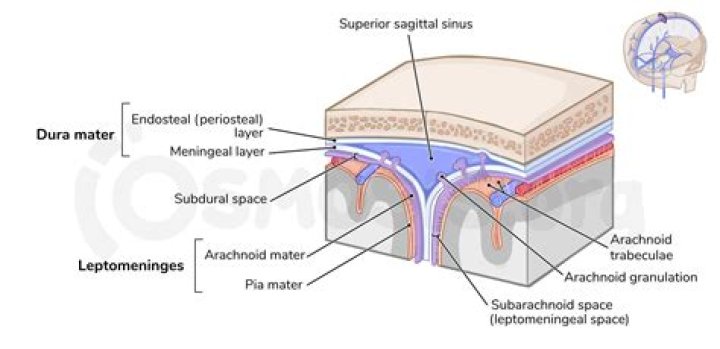
Cerebral Circulation The major dural venous sinuses include the superior and inferior sagittal sinuses, the straight sinus, the superior and inferior petrosal sinuses, the occipital sinus, the transverse sinus, and the sigmoid sinuses (Fig. 5).
What are the four regions of the dura mater?
The spinal cord and spine are divided into 4 regions from top to bottom: cervical, thoracic, lumbar, and sacral.What are dural reflections?
Dural reflections refer to places where two face-to-face meningeal layers descend into the cranial cavity to form the septa that compartmentalize the brain. The two main dural reflections are the falx cerebri and the tentorium cerebelli.
How many dural venous sinuses are there?
Unlike most veins of the body, the dural venous sinuses do not have valves. There are eleven venous sinuses in total. The straight, superior, and inferior sagittal sinuses are found in the falx cerebri of the dura mater. They converge at the confluence of sinuses (overlying the internal occipital protuberance).
How are dural reflections formed?
These are the infoldings formed by the inner meningeal layer reflecting away from the fixed periosteral dural layer. Falx cerebri (Cerebral falx): Extends from crista galli of ethmoid bone anteriorly to inner occipital protuberance posteriorly and projects over longitudinal cerebral fissure.
What are grooves of dura mater sinuses at occipital bone?
571) consists of several interlacing venous channels between the layers of the dura mater over the basilar part of the occipital bone, and serves to connect the two inferior petrosal sinuses. It communicates with the anterior vertebral venous plexus. Emissary Veins (emissaria).What is the great vein of Galen?
The great cerebral vein is one of the large blood vessels in the skull draining the cerebrum of the brain. It is also known as the “vein of Galen”, named for its discoverer, the Greek physician Galen. However, it is not the only vein with this eponym.
What is the dura of the brain?The dura mater often gets referred to as merely the dura. It is one of the layers of connective tissue that make up the meninges of the brain (pia, arachnoid, and dura, from inside to outside). It is the outermost layer of the three meninges that surround and protect the brain and spinal cord.
Article first time published onWhat is the dura?
(DER-uh MAY-ter) The tough outer layer of tissue that covers and protects the brain and spinal cord and is closest to the skull. The dura mater is one of the three layers that form the meninges.
What is the dura in the spine?
The dura is a thin layer of tissue that covers and protects the spinal cord. It lies in between the spine (the bone) and the spinal cord (nerve tissue). Durotomy, which is an incision in the dura, can be a planned part of the surgical technique.
How many sinuses are in your brain?
There are four paranasal sinuses, each corresponding with the respective bone from which it takes its name: maxillary, ethmoid, sphenoid, and frontal.
Where are bridging veins?
Bridging veins are veins in the subarachnoid space that puncture the dura mater and empty into the dural venous sinuses.
What is the arachnoid villi?
Arachnoid granulations or villi are growths of arachnoid membrane into the dural sinuses, through which the CSF enters the venous system from the subarachnoid space. 1. Arachnoid villi are microscopic, whereas arachnoid granulations represent distended villi and are visible to the naked eye.
Why is it called dura mater?
The name dura mater derives from the Latin for tough mother (or hard mother), a loan translation of Arabic أم الدماغ الصفيقة (umm al-dimāgh al-ṣafīqah), literally ‘thick mother of the brain’, matrix of the brain, and is also referred to by the term “pachymeninx” (plural “pachymeninges”).
What is Dural?
Medical Definition of dural : of or relating to the dura mater.
Does pia mater contain CSF?
Function. In conjunction with the other meningeal membranes, pia mater functions to cover and protect the central nervous system (CNS), to protect the blood vessels and enclose the venous sinuses near the CNS, to contain the cerebrospinal fluid (CSF) and to form partitions with the skull.
Is the subarachnoid space real or potential?
Comparison of Cranial and Spinal MeningesCranialSpinalSubdural SpacePotential SpacePotential SpaceArachnoidAttached to inner surface of duraAttached to inner surface of duraSubarachnoid SpaceReal Space – CSFReal Space – CSF
What is the subdural space?
The subdural space is a potential intracranial space situated between the arachnoid and dura. Fluid can collect in the subdural space and in the subarachnoid space.
Is the dura mater pain sensitive?
In this observational study, we confirmed that dura of the skull base and dura of the falx cerebri are sensitive to pain and that their mechanical stimulation induced pain mainly referred in the sensory territories of the V1 and V3 divisions of the trigeminal nerve.
How many veins are there in brain?
The brain has two main networks of veins: an exterior or superficial network, on the surface of the cerebrum that has three branches, and an interior network. These two networks communicate via anastomosing (joining) veins.
Why dural venous sinuses are valveless?
Dural venous sinuses are venous channels located intracranially between the two layers of the dura mater (endosteal layer and meningeal layer) and can be conceptualised as trapped epidural veins. … Furthermore, they are valveless, allowing for bidirectional blood flow from and into intracranial veins.
What are the venous sinuses?
venous sinus, in human anatomy, any of the channels of a branching complex sinus network that lies between layers of the dura mater, the outermost covering of the brain, and functions to collect oxygen-depleted blood. Unlike veins, these sinuses possess no muscular coat.
Which cistern does great cerebral vein occupy?
The great cerebral vein traverses the quadrigeminal cistern of the brain, which is why this area is sometimes referred to as Galen’s cistern. Within the cistern, the vein takes a posterior course, curving around the inferior aspect of the splenium of corpus callosum and reaching its posterior side.
What is Diploic vein?
The diploic veins are large, thin-walled valveless veins that channel in the diploë between the inner and outer layers of the cortical bone in the skull. They are lined by a single layer of endothelium supported by elastic tissue. … The diploic veins drain this area into the dural venous sinuses.
What is a Galen aneurysm?
Listen. Vein of Galen aneurysm is a rare form of arteriovenous malformation in which the embryonic precursor to the vein of Galen, a vein at the base of the brain, dilates causing too much blood to rush to the heart. This can lead to rapid heart failure.
What do dural venous sinuses carry?
The dural venous sinuses are spaces between the endosteal and meningeal layers of the dura. They contain venous blood that originates for the most part from the brain or cranial cavity. The sinuses contain an endothelial lining that is continuous into the veins that are connected to them.
What is dural venous sinus thrombosis?
A dural sinus thrombosis is the occlusion of a dural sinus by a blood clot (or thrombus). Because of this occlusion, blood flowing out of the brain is backed up, and the brain tissue becomes congested. As a result, both ischemia and hemorrhage may occur.
Does the dural sinus contain CSF?
NameDrains toPosteriorOccipital sinusConfluence of SinusConfluence of sinusesRight and Left transverse sinusesLateral
What are the 3 layers of the brain?
Three layers of membranes known as meninges protect the brain and spinal cord. The delicate inner layer is the pia mater. The middle layer is the arachnoid, a web-like structure filled with fluid that cushions the brain. The tough outer layer is called the dura mater.