What are somites function

Christopher Lucas
Published Mar 01, 2026
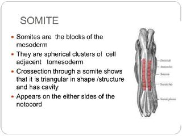
Somites are literally the building blocks of the vertebrate body plan; they are essential for segmentation, bone and musculature development, as well as creating a template for the nervous system.
How many somites do humans have?
In humans 42-44 somite pairs 9 – 13 are formed along the neural tube. These range from the cranial region up to the embryo’s tail. Several caudal somites disappear again, which is why only 35-37 somite pairs can be counted in the end.
What is a somite in medicine?
[so´mīt] one of the paired segments along the neural tube of a vertebrate embryo, formed by transverse subdivision of the thickened mesoderm next to the midplane, that develop into the vertebral column and muscles of the body.
How do somites differentiate?
Cells situated ventromedially in a somite differentiate into the sclerotome, which gives rise to cartilage, while the other part of the somite differentiates into dermomyotome which gives rise to muscle and dermis.What tissues are formed by somites?
Somites give rise to the cells that form the vertebrae and ribs, the dermis of the dorsal skin, the skeletal muscles of the back, and the skeletal muscles of the body wall and limbs.
What is somites in biology?
somite, in embryology, one of a longitudinal series of blocklike segments into which the mesoderm, the middle layer of tissue, on either side of the embryonic spine becomes divided. … The term somite is also used more generally to refer to a body segment, or metamere, of a segmented animal.
Are somites mesenchyme?
The outer cells undergo a mesenchymal–epithelial transition to form an epithelium around each somite. The inner cells remain as mesenchyme.
Are somites epithelial?
Somites bud off sequentially and rhythmically from the mesenchymal ‘paraxial’ mesoderm, arising as pairs of epithelial spheres that flank the neural tube and accumulate in a progressive A-P direction. … Together with cells from the midline notochord, the sclerotome differentiates into the vertebral column.What are occipital Myotomes?
The occipital myotomes (4, light green) mainly form the pharynx (throat) and upper or anterior neck musculature, including the tongue muscles. They are also responsible for the musculature in the occipital head region.
How is Chordamesoderm formed?Axial mesoderm, or chordamesoderm, is the mesoderm in the embryo that lies along the central axis under the neural tube. starts as the notochordal process, whose formation finishes at day 20 in humans. … The notochord will form the nucleus pulposus of intervertebral discs.
Article first time published onHow many somites are in a chick embryo?
In the chick embryo, a pair of somite forms every 90 min at 37 °C and a total of 52 somites pairs are formed during the somitogenesis process which lasts from day 1 to day 5 of development. Somitogenesis can be subdivided into three major phases.
What is occipital somites?
Occipital somites (1-5 in human) fuse at an early stage of embryonic development and do not contribute to segmented skeletal structures. They incorporate into the occipital area of embryonic skull, give rise to the tongue muscles and also condense to contribute to the basi-occipital and exo-occipital cartilages.
What is primitive streak?
The primitive streak is a structure that forms in the blastula during the early stages of avian, reptilian and mammalian embryonic development. It forms on the dorsal (back) face of the developing embryo, toward the caudal or posterior end.
What is neural crest?
The neural crest is a transient embryonic structure in vertebrates that gives rise to most of the peripheral nervous system (PNS) and to several non-neural cell types, including smooth muscle cells of the cardiovascular system, pigment cells in the skin, and craniofacial bones, cartilage, and connective tissue.
How many segments or somites make up the spinal cord?
Dermatomes of the head, face, and neck. The spinal cord has 31 segments, each with a pair (right and left) of ventral (anterior) and dorsal (posterior) nerve roots that innervate motor and sensory function, respectively.
How does the blastocyst develop?
In humans, blastocyst formation begins about 5 days after fertilization when a fluid-filled cavity opens up in the morula, the early embryonic stage of a ball of 16 cells. … About seven days after fertilization, the blastocyst undergoes implantation, embedding into the endometrium of the uterine wall.
Which gives rise to skeletal muscle?
The ventral part of the somite, the sclerotome, will contribute the cartilage and bone of the vertebral column and ribs, whereas the dorsal part of the somite, the dermomyotome, as its name implies, gives rise to the overlying derm of the back and to the skeletal muscles of the body and limbs.
What general feature's of vertebrates is are associated with somites?
Segmented structures composed of repetitive units, called somites, that arise transiently during embryogenesis are a key feature of the vertebrate body plan. The somites lie laterally to the notochord, and a spinal nerve forms a segmental unit assigned to somitic derivatives in the trunk [1, 2].
How many somites can you expect to see in the 24 hour chick embryo?
Fig. 40. Dextral view of entire chick embryo of 41 somites (about four days incubation). Note that in the 24 hour chick, Hensen’s node is located further caudally and the primitive streak is present only at the posterior end of the embryo.
Is a notochord a spine?
A notochord does not become the spine, but rather fills the spaces between vertebrae in some animals.
What is mesenchyme tissue?
Mesenchyme is a type of animal tissue comprised of loose cells embedded in a mesh of proteins and fluid, called the extracellular matrix. … Mesenchyme directly gives rise to most of the body’s connective tissues, from bones and cartilage to the lymphatic and circulatory systems.
What is the neural plate?
The neural plate is a cohesive structure whose cells are linked together by junctional complexes. There are structural changes that occur within neuroepithelial cells of the neural plate that contribute to the formation of the neural groove and then the neural tube.
What is pharyngeal arch?
Anatomy: Pharyngeal arches are paired structures that grow on either side of the future head and neck of the developing embryo and fuse at the centerline. … Pharyngeal arches produce the cartilage, bone, nerves, muscles, glands, and connective tissue of the face and neck.
What is Myotomes and Dermatomes?
What are Myotomes and Dermatomes? A group of muscles that is innervated by the motor fibers that stem from a specific nerve root is called a myotome. An area of the skin that is innervated by the sensory fibers that stem from a specific nerve root is called a dermatome.
What is L1 myotome?
The lumbar and sacral myotomes (L1-S3) are tested with the patient lying supine. These are tested with movements of the hip, knee, ankle, intertarsal, and metatarsophalangeal joints. Movements for lumbar and sacral myotome testing.
What structure do the Chordamesoderm cells produce?
The next cells involuting into the embryo through the dorsal blastopore lip are called the chordamesoderm cells. These cells will form the notochord, a transient mesodermal “backbone” that plays an important role in distinguishing and patterning the nervous system.
What are the other structures formed by the Blastoderm present external to the embryo?
Serosal Formation Only blastoderm cells destined to form the embryo coalesce to form the germ anlage, which later develops into the germ band. … In addition to the serosa, a second protective membrane, the amnion, forms later from the cells immediately adjacent to the germ anlage.
What will become of the Archenteron in the developing frog embryo?
During gastrulation, the archenteron develops into the digestive tube, with the blastopore developing into either the mouth (protostome) or the anus (deuterostome).
What is frog gastrulation?
Gastrulation in the process of highly integrated cell and tissue migrations of prospective endodermal and mesodermal areas to their definite positions into the interior of the embryo. There occur three types of morphogenetic movements in amphibian gastrulation. …
What is the organ that will develop first in the chick embryo?
The chick embryo is ideal for studying the early development of the heart, the first functioning organ in the embryo. A major advantage is that the chick develops ex utero in an egg, which allows easy accessibility during all stages of development post-laying.
What is gastrulation biology?
Gastrulation is defined as an early developmental process in which an embryo transforms from a one-dimensional layer of epithelial cells (blastula) and reorganizes into a multilayered and multidimensional structure called the gastrula.