Is there a vertebral vein

Mia Kelly
Published Feb 23, 2026
The vertebral vein. (Vertebral labeled at upper left and center right.) The vertebral vein is formed in the suboccipital triangle, from numerous small tributaries which spring from the internal vertebral venous plexuses and issue from the vertebral canal above the posterior arch of the atlas.
How many vertebral veins are there?
the two vertebral arteries. the two vertebral veins. the anterior spinal artery. the two posterior spinal arteries.
Is there a left and right vertebral vein?
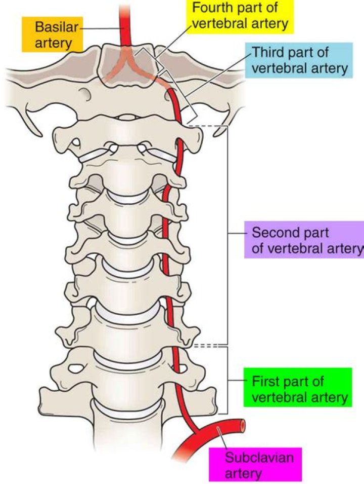
The vertebral arteries in the neck supply blood to the brain and spine. The name vertebral refers to the arteries’ location along the vertebrae, the bones of the spine. You have a left vertebral artery and a right vertebral artery that run through the spinal column.
Where does the vertebral vein supply blood to?
The Vertebral and Internal Carotid Arteries provide blood to the brain. These arteries give off branches that form a circle in the region of the pituitary gland. If the other two arteries are blocked, the blood vessels in the Circle of Willis provide an alternate way to feed blood to the brain.Which veins drain vertebral bodies?
Intervertebral veinsLatinvena intervertebralisAnatomical terminology
What does vertebral vein drain?
The function of the vertebral vein is to drain the venous blood from the cervical spine, prevertebral and suboccipital muscles. It terminates low in the neck by emptying into the brachiocephalic vein.
Is the spinal cord a vein?
Spinal veinsFMA71580Anatomical terminology
What does the subclavian vein drain?
The primary function of the subclavian vein is to drain deoxygenated blood from the upper region of the body—including the arms and the shoulder areas—and transport it back to the heart. 6 Another important function of the subclavian is to collect lymph fluid from the lymphatic system from the internal jugular vein.What is the longest vein in the body?
Great Saphenous Vein (GSV) – The GSV is the large superficial vein of the leg and the longest vein in the entire body. It can be found along the length of the lower limb, returning blood from the thigh, calf, and foot to the deep femoral vein at the femoral triangle. The femoral triangle is located in the upper thigh.
Where are your arteries in your back?The lumbar arteries are arteries located in the lower back or lumbar region. The lumbar arteries are in parallel with the intercostals. They are usually four in number on either side, and arise from the back of the aorta, opposite the bodies of the upper four lumbar vertebrae.
Article first time published onCan you get varicose veins in your back?
Varicose veins usually develop on the legs, either on the back of your calf or on the inside of your leg. However, they can also sometimes occur in other parts of your body, such as your: gullet (oesophagus) womb (uterus)
What vertebrae is the heart at?
The highest point of the base of the heart varies in level from the lower third of the fourth to the upper third of the eighth thoracic vertebrae, with the average position being at a level between the middle and lower thirds of the sixth thoracic vertebra, as reported by Eycleshymer and Schoemaker.
What happens if the vertebral artery is blocked?
These arteries supply blood to the brainstem and the cerebellum. Like carotid artery stenosis, vertebral artery stenosis is highly dangerous and can prevent oxygen from reaching the brain. When the brain doesn’t get enough oxygen, a stroke, or even death, can occur.
Can vertebral artery cause vertigo?
OVERVIEW. In a vertebral artery dissection, blood enters between layers of the vertebral artery, resulting in diminished blood flow. This can cause a stroke, dizziness and vertigo, visual disturbances, and numerous other neurological disturbances.
Is your neck connected to your spine?
The neck is connected to the upper back through a series of seven vertebral segments. The cervical spine has 7 stacked bones called vertebrae, labeled C1 through C7. The top of the cervical spine connects to the skull, and the bottom connects to the upper back at about shoulder level.
How do you treat spinal cord compression?
- Medicines may include nonsteroidal anti-inflammatory drugs (NSAIDs) that relieve pain and swelling, and steroid injections that reduce swelling.
- Physical therapy may include exercises to strengthen your back, abdominal, and leg muscles.
Where does external vertebral venous plexus drain?
After receiving blood from the region of the vertebral column, the internal venous plexus drains into the external venous plexus via the intervertebral veins. In turn, the external one drains into the vertebral veins of the neck and the segmental veins of the trunk.
Where does the spinal cord end?
The spinal cord is an extension of the central nervous system (CNS), which consists of the brain and spinal cord. The spinal cord begins at the bottom of the brain stem (at the area called the medulla oblongata) and ends in the lower back, as it tapers to form a cone called the conus medullaris.
Why is lumbar puncture done?
A lumbar puncture can help diagnose serious infections, such as meningitis; other disorders of the central nervous system, such as Guillain-Barre syndrome and multiple sclerosis; or cancers of the brain or spinal cord.
What is Brown Séquard syndrome?
Brown-Séquard syndrome is a rare spinal disorder that results from an injury to one side of the spinal cord in which the spinal cord is damaged but is not severed completely. It is usually caused by an injury to the spine in the region of the neck or back.
What is vertebral plexus?
The vertebral venous plexus, also called the Batson plexus refers to veins located extradurally within the spinal canal (internal plexus), surrounding the vertebral column (external plexus) as well as horizontal basivertebral veins that run through the vertebral bodies (basivertebral plexus).[1][2] These veins comprise …
What is right subclavian vein?
The right subclavian vein is joined by the right lymphatic duct at the right venous angle, which drains lymph from the right upper quadrant of the body (right side of the head, neck, thorax, right upper extremity).
What is vertebral column?
(ver-TEE-brul KAH-lum) The bones, muscles, tendons, and other tissues that reach from the base of the skull to the tailbone. The vertebral column encloses the spinal cord and the fluid surrounding the spinal cord. Also called backbone, spinal column, and spine.
What vein enter the vertebral veins of the left side in?
The inferior vena cava (IVC) is a large retroperitoneal vessel formed by the confluence of the right and left common iliac veins. Anatomically this usually occurs at the L5 vertebral level.
What does saphenous mean?
Definition of saphenous : of, relating to, associated with, or being either of the two chief superficial veins of the leg saphenous nerve.
What is the strongest vein in the body?
The two largest veins in the body are the superior vena cava, which carries blood from the upper body directly to the right atrium of the heart, and the inferior vena cava, which carries blood from the lower body directly to the right atrium.
Where is the best place to find a vein?
Use veins on top of the hand, top of the forearm, or inside the elbow. Veins inside the elbow are large but hard to reach by yourself. It is easier to reach the smaller veins on top of your hand. Feel veins to see how big they are and in which direction they go.
What happens if the subclavian vein is blocked?
This tissue causes the vein to narrow and restrict blood flow, leading to the formation of blood clots. Left untreated, axillo-subclavian vein thrombosis can cause: Arm pain and fatigue. Arm swelling.
Is subclavian vein a deep vein?
In the upper extremity the deep veins include the paired radial veins, paired ulnar veins, paired brachial veins, axillary vein, and subclavian vein.
Where does the subclavian vein lie?
The subclavian vein is the major vein of the arm, shoulder and neck. Its name means ‘under the clavicle’, due to the course it takes when entering the thorax.
What causes vertebral artery blockage?
Atherosclerosis or “hardening of the arteries” is the main cause of vertebrobasilar disease. The narrowing of the vertebral or basilar arteries caused by atherosclerosis creates vertebrobasilar insufficiency (VBI), or an insufficient delivery of blood flow to the posterior structures of the brain.