How many mm is an ECG box

Emma Valentine
Published Feb 13, 2026
Electrocardiogram waves, intervals, and segments. On standard calibration, each large box has sides of 0.5 cm. On the horizontal axis, each large box represents 0.2 seconds, and each smaller box represents 0.04 seconds. On the vertical axis, each small box is 1 mm in height; 10 mm = 1 mV.
What is 1 mm on an ECG?
Each small square is 1 mm in length and represents 0.04 seconds. Each larger square is 5 mm in length and represents 0.2 seconds.
What are ECG measurements?
An electrocardiogram (ECG or EKG) is a test that checks how your heart is functioning by measuring the electrical activity of the heart. With each heart beat, an electrical impulse (or wave) travels through your heart.
How are ECG boxes measured?
- 1 SMALL square = 0.04 seconds.
- 5 SMALL squares = 1 LARGE square = 0.2 seconds.
- 5 LARGE squares = 1 second.
- ECG rhythm strip: = 250 SMALL squares = 50 LARGE squares = 10 seconds.
- To calculate beats per minute (bpm): 1500 SMALL squares = 300 LARGE squares = 1 minute.
How much is each little box on ECG?
As a result, each 1 mm (small) horizontal box corresponds to 0.04 sec (40 ms), with heavier lines forming larger boxes that include five small boxes and hence represent 0.20 sec (200 ms) intervals.
How many squares is 1 second in ECG?
Each ECG is divided by large boxes and small boxes to help measure times and distances. Each large box represents 0.20 seconds, and there are five small boxes in each large box, thus each small box is equivalent to 0.04 seconds.
How many boxes is 120 ms on ECG?
3 small squares (120ms) is abnormal, not the upper limit of nor- mal. If necessary, use a magnifying glass.
What does V1 V2 V3 mean in ECG?
The areas represented on the ECG are summarized below: V1, V2 = RV. V3, V4 = septum. V5, V6 = L side of the heart. Lead I = L side of the heart.What is the normal ECG rate?
A normal ECG is illustrated above. Note that the heart is beating in a regular sinus rhythm between 60 – 100 beats per minute (specifically 82 bpm). All the important intervals on this recording are within normal ranges.
How do you report a normal ECG?- Confirm correct patient details.
- Rate.
- Rhythm.
- Cardiac axis.
- P waves, Q waves & QRS complexes.
- ST segments & T waves.
- QT interval.
- Putting it all together.
How many small boxes is normal QT interval?
QT interval = about 10 small squares = 0.4 seconds.
How many boxes is a normal QRS?
This measurement should be 0.12-0.20 seconds, or 3-5 small squares in duration. The second measurement is the width of the QRS which should be less than 3 small squares, or less than 0.12 seconds in duration.
How many boxes is a normal QT interval?
For normal rates, QT < . 4 seconds (2 large boxes). “QT prolongation” (too long) can lead to a refractory form of ventricular tachycardia called torsades de pointes. Figure 27: The QT interval is greater than half the preceding RR interval.
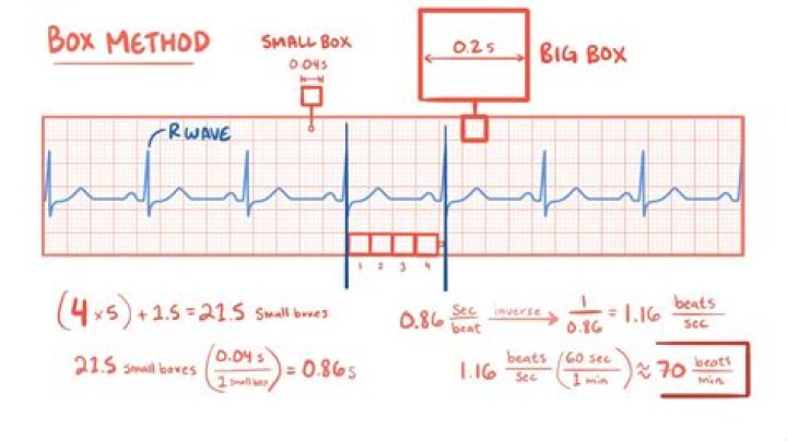
What is the 300 rule for ECG?
The 300 Method: Count the number of large boxes between 2 successive R waves and divide by 300 to obtain heart rate. 4. The 1500 Method: Count the number of small boxes between two successive R waves and divide this number into 1500 to obtain heart rate. This works well for faster heart rates.
What are the 4 lethal heart rhythms?
You will need to be able to recognize the four lethal rhythms. Asystole, Ventricle Tachycardia (VT), Ventricle Fibrillation (VF), and Polymorphic Ventricle Tachycardia (Torsade de pointes). Use this study guide and other resource books to review ECG interpretation.
When is an 18 lead right sided ECG used?
Conclusion. The diagnosis of STEMI by synthesized 18-lead ECG is useful to identify the site of infarction in patients with infarction of the right ventricular wall (supplied by the RCA) or posterior wall of the left ventricle (supplied by the LCX), which often fail to be diagnosed by the standard 12-lead ECG.
How many small boxes fit in a large box ECG?
On the horizontal axis, each large box represents 0.2 seconds at a typical paper speed of 25 mm per second, which is then divided into five smaller boxes that each represent 0.04 seconds.
How many small squares is ST elevation?
An ST elevation is considered significant if the vertical distance inside the ECG trace and the baseline at a point 0.04 seconds after the J-point is at least 0.1 mV (usually representing 1 mm or 1 small square) in a limb lead or 0.2 mV (2 mm or 2 small squares) in a precordial lead.
How many boxes are in P wave?
This rhythm is actually an accelerated idioventricular rhythm, or slow ventricular tachycardia. The atrial rate is indicated by the P waves. There are almost exactly five large boxes between P waves, indicating an atrial rate of 60 bpm.
What is abnormal ECG?
An abnormal ECG can mean many things. Sometimes an ECG abnormality is a normal variation of a heart’s rhythm, which does not affect your health. Other times, an abnormal ECG can signal a medical emergency, such as a myocardial infarction /heart attack or a dangerous arrhythmia.
Can ECG detect heart blockage?
However, it does not show whether you have asymptomatic blockages in your heart arteries or predict your risk of a future heart attack. The resting ECG is different from a stress or exercise ECG or cardiac imaging test.
What is a 3 lead ECG used for?
3-lead ECGs are used most often for recording a 24-hour reading. A 24-hour reading is a frequently used tool for the diagnosis of heart problems and is reimbursed as a long-term reading.
What does inverted T wave mean on ECG?
Inverted T waves. Ischemia: Myocardial ischemia is a common cause of inverted T waves. Inverted T waves are less specific than ST segment depression for ischemia, and do not in and of themselves convey a poor prognosis (as compared to patients with an acute coronary syndrome and ST segment depression).
Are V1 V6 unipolar or bipolar?
The electrode leads each have a name. The bipolar extremity leads are called I, II and III. The unipolar extremity leads are called avR, avL and avF, and the chest leads are called V1–V6.
What should my heartbeat look like?
The average healthy adult will have a resting heart rate of 60 bpm or higher. Although in clinical practice, the resting heart rate between 60 and 100 bpm is considered to be normal, people with a resting heart rate higher than 80 bpm could have an increased risk of developing cardiovascular disease.
Why do we use lead 2 in ECG?
The most commonly used lead is lead II – a bipolar lead with electrodes on the right arm and left leg. This is the most useful lead for detecting cardiac arrhythmias as it lies close to the cardiac axis (the overall direction of electrical movement) and allows the best view of P and R waves.
What is P in ECG report?
The P wave and PR segment is an integral part of an electrocardiogram (ECG). It represents the electrical depolarization of the atria of the heart. It is typically a small positive deflection from the isoelectric baseline that occurs just before the QRS complex.
How many small boxes wide would a normal QRS wave measure?
1 small square on an ECG trace (at 25 mm/s speed) = 0.04 s. The P wave 0.08–0.11 seconds (2–3 small squares) PR interval 0.11–0.20 seconds (3–5 small squares) QRS complex 0.06–0.11 seconds (1.5–2.5 small squares)