How is CSF fluid produced

Victoria Simmons
Published Feb 20, 2026
CSF is produced by specialised ependymal cells in the choroid plexus of the ventricles of the brain, and absorbed in the arachnoid granulations. … It fills the ventricles of the brain, cisterns, and sulci, as well as the central canal of the spinal cord.
How is CSF produced and circulated?
According to the traditional understanding of cerebrospinal fluid (CSF) physiology, the majority of CSF is produced by the choroid plexus, circulates through the ventricles, the cisterns, and the subarachnoid space to be absorbed into the blood by the arachnoid villi.
Is CSF constantly produced?
The CSF is continually produced, and all of it is replaced every six to eight hours. The fluid is eventually absorbed into the veins; it leaves the cerebrospinal spaces in a variety of locations, including spaces around the spinal roots and the cranial nerves.
How is CSF produced and reabsorbed?
The CSF from the subarachnoid space is eventually reabsorbed through outpouchings into the superior sagittal sinus (SSS) known as the arachnoid granulations. Arachnoid granulations act as an avenue for CSF reabsorption into the blood circulation through a pressure-dependent gradient.Which cell produces CSF?
The choroid plexus is a complex network of capillaries lined by specialized cells and has various functions. One of the primary functions is to produce cerebrospinal fluid (CSF) via the ependymal cells that line the ventricles of the brain.
Where is CSF produced and reabsorbed?
The traditional hypothesis of cerebrospinal fluid (CSF) hydrodynamics presumes that CSF is primarily produced in the choroid plexus (CP), then flows from the ventricles into the subarachnoid spaces, and mainly reabsorbed in the arachnoid granulations.
How can I increase my CSF flow?
These techniques include massage and manipulation of the spine. Walking, stretching, cycling, heat, and yoga may all help SFF.
How does CSF enter the subarachnoid space?
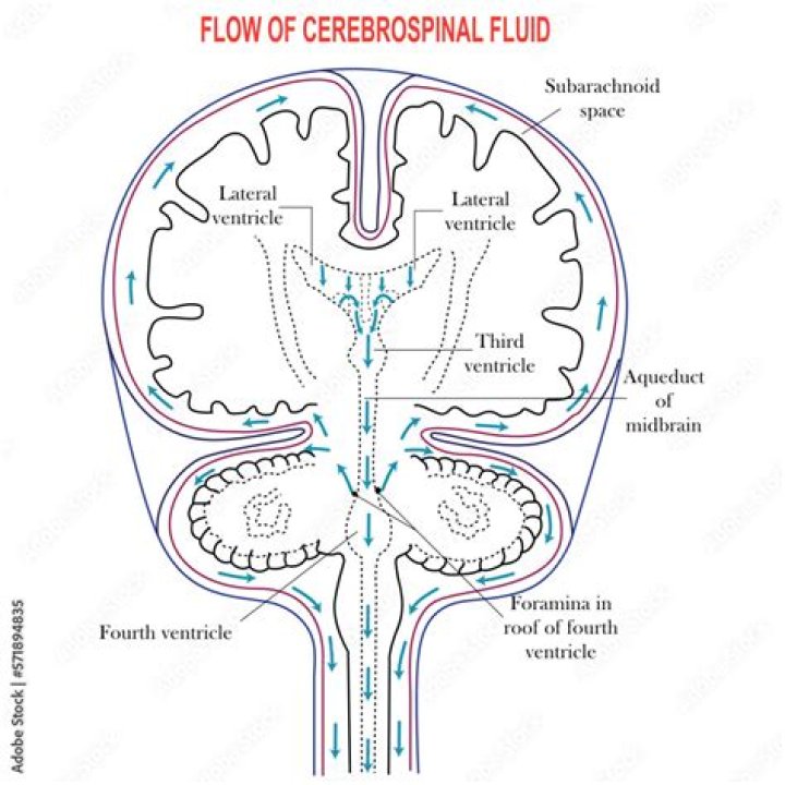
CSF flows from the lateral ventricle to the third ventricle through the interventricular foramen (also called the foramen of Monro). … CSF then flows into the subarachnoid space through the foramina of Luschka (there are two of these) and the foramen of Magendie (only one of these).How does CSF get into the spinal cord?
The majority of CSF exits from the fourth ventricle into the subarachnoid space; a small amount may enter the central canal of the spinal cord. In people, CSF enters the subarachnoid space through the lateral apertures (foramina of Luschka) and the median aperture (foramen of Magendie) of the fourth ventricle.
How much CSF is produced per hour?In normal adults, the CSF volume is 90 to 200 mL [1]; approximately 20 percent of the CSF is contained in the ventricles; the rest is contained in the subarachnoid space in the cranium and spinal cord. The normal rate of CSF production is approximately 20 mL per hour.
Article first time published onWhat color is CSF?
Color of the fluid—normal is clear and colorless. Changes in the color of the CSF are not diagnostic but may point to additional substances in the fluid. Yellow, orange, or pink CSF may indicate the breakdown of blood cells due to bleeding into the CSF or the presence of bilirubin.
How do you reduce CSF production?
Acetazolamide (ACTZ), a carbonic anhydrase inhibitor, has been shown to decrease cerebrospinal fluid (CSF) production in both in vivo and in vitro animal models.
How does CSF work?
Cerebrospinal Fluid (CSF) flows through the four ventricles and then flows between the meninges in an area called the subarachnoid space. CSF cushions the brain and spinal cord against forceful blows distributes important substances and carries away waste products.
How do ependymal cells produce CSF?
The layer of ependymal-derived cells surrounding the blood vessels of the choroid plexus functions mainly to produce CSF. … Because the junctions between the ependymal cells are loose, CSF is able to diffuse from the ventricles into the central nervous system.
How does the choroid plexus produce CSF?
CSF is formed as plasma is filtered from the blood through the epithelial cells. Choroid plexus epithelial cells actively transport sodium ions into the ventricles and water follows the resulting osmotic gradient. … Fluid filters through these cells from blood to become cerebrospinal fluid.
What is the normal composition of CSF?
Normal Results CSF total protein: 15 to 60 mg/100 mL. Gamma globulin: 3% to 12% of the total protein. CSF glucose: 50 to 80 mg/100 mL (or greater than two thirds of blood sugar level) CSF cell count: 0 to 5 white blood cells (all mononuclear), and no red blood cells.
What affects CSF production?
CSF circulation from sites of secretion to sites of absorption largely depends on the arterial pulse wave. Additional factors such as respiratory waves, the subject’s posture, jugular venous pressure and physical effort also modulate CSF flow dynamics and pressure.
What happens if you have too much CSF?
The body typically produces enough CSF each day and absorbs the same amount. However, when the normal flow or absorption of CSF is blocked it can result in a buildup of CSF. The pressure from too much CSF can keep the brain from functioning properly and cause brain damage and even death.
What two things maintain CSF pressure?
Undoubtedly CSF secretion, absorption and drainage are important aspects of brain fluid homeostasis in maintaining a stable ICP.
How does CSF leave the subarachnoid space?
The CSF exits the subarachnoid space by diffusing through the walls of arachnoid granulations. The arachnoid granulations provide a valvular mechanism for the flow of CSF, which allows the inflow of CSF into the bloodstream without permitting the backflow of blood into the CSF.
Where does CSF drain?
CSF gets drained into the superior sagittal venous sinus through the arachnoid villi, small protrusions of arachnoid matter into the venous sinus. Physiologically, the pressure of CSF within the subarachnoid space is greater than that within the venous sinus. Hence, the CSF will drain into the venous sinuses.
What regulates CSF pressure?
The secretion and composition of the CSF is tightly regulated by the CPs, which are complex structures comprised of a plexus of fenestrated capillaries surrounded by a layer of cuboidal epithelial cells, with an intervening stromal space between these two components (Fig. 1D).
What is the pH of CSF?
In the arterial blood the pH was 7.20, the PaCO2 20.0 mm. of mercury, and the bicarbonate concentration 7.4 mEq. per liter. Contrasting with this blood acidosis, the pH of the cerebrospinal fluid was 7.45, the PCO2 30 mm.
Is CSF test painful?
During the procedure: You will lie on your side or sit on an exam table. A health care provider will clean your back and inject an anesthetic into your skin, so you won’t feel pain during the procedure.
Is CSF fluid sticky?
Unlike mucus, which is thick and sticky, CSF is clear and watery. Compared with mucus, CSF also has a high concentration of glucose. Checking the glucose levels in nasal discharge can help determine whether it contains CSF.
How do I know if my fluid is CSF?
Your middle ear fluid may be tested to check for CSF . CT myelography. This test is considered the gold standard for diagnosing and locating CSF leaks. It uses a CT scan and a contrast dye to locate CSF leaks anywhere in the skull base.
What foods increase CSF?
five portions of fruits and vegetables per day. carbohydrates from foods such as brown rice, potatoes, cereals and whole wheat pasta. protein from foods such as oily fish, eggs and meat.
Does exercise increase CSF flow?
Cerebral blood flow (CBF) increases during exercise, but its impact on cerebrospinal fluid (CSF) flow remains unknown. This study investigated CBF and CSF flow dynamics during moderate-intensity rhythmic handgrip (RHG) exercise in young healthy men and women.
Does caffeine increase CSF?
The results of this study show that the long-term consumption of caffeine can induce ventriculomegaly, which is mediated in part by increased production of CSF. Moreover, adenosine receptor signaling appears to regulate the production of CSF by controlling the expression of Na+, K+-ATPase and CBF.