How does MS appear on MRI

Robert Spencer
Published Feb 20, 2026
MS-related lesions appear on MRI images as either bright or dark spots, depending on the type of MRI used. This imaging technique is useful because it shows active inflammation and helps doctors determine the age of the lesions. Also, some specific types of lesion can indicate a flare-up of MS or damage in the brain.
Can an MRI scan rule out MS?
MRI is considered the best test to help diagnose MS. However, 5% of people with MS do not have abnormalities detected on MRI; thus, a “negative” scan does not completely rule out MS. In addition, some common changes of aging may look like MS on a MRI.
What are usually the first signs of MS?
- vision problems.
- tingling and numbness.
- pains and spasms.
- weakness or fatigue.
- balance problems or dizziness.
- bladder issues.
- sexual dysfunction.
- cognitive problems.
Do MS lesions show up on MRI without contrast?
MS patients can be effectively monitored without the use of contrast agents. Researchers assessed 507 follow-up MR images for new or enlarged lesions. The 3T MRI results did not differ significantly between contrast-enhanced and non-enhanced images.Is it possible to have MS without lesions?
About 5 percent of people who are confirmed to have MS do not initially have brain lesions evidenced by MRI. However, the longer a person goes without brain or spinal cord lesions on MRI, the more important it becomes to look for other possible diagnoses.
What can an MRI not detect?
MRI can be used to view arteries and veins. Standard MRI can’t see fluid that is moving, such as blood in an artery, and this creates “flow voids” that appear as black holes on the image. Contrast dye (gadolinium) injected into the bloodstream helps the computer “see” the arteries and veins.
Does MRI show early MS?
The magnetic resonance imaging (MRI) may show areas of abnormality that suggest MS, though the MRI in and of itself does not make the diagnosis. Spinal fluid testing may show that the immune system is active in and around the brain and spinal cord, supporting the diagnosis. Evoked potentials may assist in diagnosis.
How accurate is MRI in diagnosing multiple sclerosis?
MRI has greater than 90% sensitivity in the diagnosis of MS; however, other white matter diseases can sometimes have a similar appearance on medical imaging.What is the McDonald criteria for MS diagnosis?
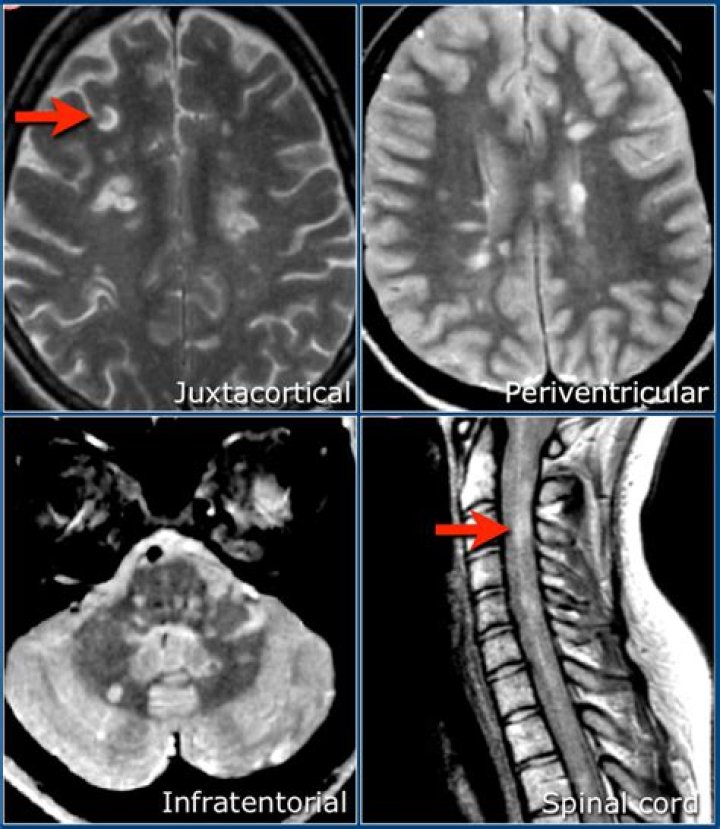
Under the McDonald Criteria (revised), an MS diagnosis is likely if myelin damage is disseminated in space, as seen in an MRI as: At least one T2 bright lesion in at least two or four CNS locations: the juxtacortical, perventricular and infratentorial areas of the brain, and the spinal cord.
Can anxiety mimic MS?Never Self-Diagnose MS From Anxiety Unfortunately, anxiety causes many of the same symptoms as the early stages of MS. MS is one of the health issues that comes up most when those with anxiety search for their symptoms online, and millions of those with anxiety convince themselves that they might have MS.
Article first time published onWhat shows up bright white on an MRI?
On a T1-weighted scans show tissues with high fat content (such as white matter) appear bright and compartments filled with water (CSF) appears dark. This is good for demonstrating anatomy.
Where does MS usually start?
Here’s where MS (typically) starts Optic neuritis, or inflammation of the optic nerve, is usually the most common, Shoemaker says. You may experience eye pain, blurred vision and headache.
Does MS show up in blood work?
No blood test can diagnose MS individually. However, the doctor may order blood testing to rule out other conditions that have similar symptoms. Blood testing can rule out the following health problems: Lyme disease.
What age does MS usually start?
Age. MS can occur at any age, but onset usually occurs around 20 and 40 years of age. However, younger and older people can be affected.
What does an MS hug feel like?
The ‘MS hug’ is symptom of MS that feels like an uncomfortable, sometimes painful feeling of tightness or pressure, usually around your stomach or chest. The pain or tightness can stretch all around the chest or stomach, or it can be just on one side. The MS hug can feel different from one person to another.
Can Covid mimic MS?
There is a proven association between HCoV and MS pathogenesis which has evolved from several experimental studies which revealed that murine coronavirus infection of susceptible mice led to an inflammatory demyelination similar to MS; coronavirus RNA sequences and its antigen were detected in demyelinating lesions [4 …
What kind of MRI can diagnose MS?
A common type of MRI for MS is a T2-weighted scan, which detects all areas of myelin damage in the brain and spinal cord. We can now use a technique called FLAIR to make it easier to spot the lesions. Doctors will also use a contrast agent called gadolinium with a T1-weighted scan to focus on newer, active lesions.
Will an MRI show nerve damage?
An MRI may be able help identify structural lesions that may be pressing against the nerve so the problem can be corrected before permanent nerve damage occurs. Nerve damage can usually be diagnosed based on a neurological examination and can be correlated by MRI scan findings.
Does MRI show all problems?
Possible findings. It is possible that an MRI may show that everything is completely normal; however, there are several things that could be seen on an MRI and this will vary depending on where in the body the scan is being done. An MRI is very good at showing up problems with soft tissues such as muscles and ligaments …
Does MRI show inflammation?
MRI allows to assess the soft tissue and bone marrow involvement in case of inflammation and/or infection. MRI is capable of detecting more inflammatory lesions and erosions than US, X-ray, or CT.
How long on average does it take to diagnose MS?
It can take a few years to make an accurate diagnosis of progressive MS because the condition usually worsens slowly.
What are MS attacks like?
Multiple sclerosis (MS) attacks can include tingling, numbness, fatigue, cramps, tightness, dizziness, and more. Multiple sclerosis (MS) is an autoimmune disorder in which your own antibodies (autoantibodies) start attacking and destroying the nerve cells of your body.
Can a neurologist diagnose MS?
To diagnose MS, your neurologist will conduct a neurological exam, which is a physical exam to determine nerve function; request MRI studies of your brain and/or spine; and conduct a spinal tap. If you are experiencing vision problems, there will be an eye exam, as well.
What kind of MRI do I need for multiple sclerosis?
A: We recommend an initial cervical and thoracic spine MRI with and without contrast along with brain MRI in patients suspected of having MS, for diagnosis, to establish disease burden, and to monitor for asymptomatic spinal cord lesions[4,5].
Can an MRI detect ALS?
Scans. Scans such as magnetic resonance imaging, or MRI, can’t directly diagnose ALS. That’s because people with the condition have normal MRI scans. But they are often used to rule out other diseases.
How can I test myself for multiple sclerosis?
There are no specific tests for MS . Instead, a diagnosis of multiple sclerosis often relies on ruling out other conditions that might produce similar signs and symptoms, known as a differential diagnosis. Your doctor is likely to start with a thorough medical history and examination.
What are the symptoms of MS in a woman?
- Vision problems. For many people, a vision problem is the first noticeable symptom of MS. …
- Numbness. …
- Fatigue. …
- Bladder problems. …
- Bowel problems. …
- Pain. …
- Cognitive changes. …
- Depression.
Can you have MS for years and not know it?
Benign MS can’t be identified at the time of initial diagnosis; it can take as long as 15 years to diagnose. The course of MS is unpredictable, and having benign MS doesn’t mean that it can’t progress into a more severe form of MS.
What are T2 lesions?
T2/FLAIR lesions can directly account for some symptoms. For example, a brainstem lesion can cause room spinning sensations and balance problems. Cervical (neck) spinal cord T2/FLAIR lesions could cause tingling and numbness in the hands and legs. Many of the lesions may not be causing obvious symptoms.
Are white spots on brain MRI normal?
White spots on a brain MRI are not always a reason for concern. There are many possible causes, including vitamin deficiencies, infections, migraines, and strokes.
What does a bright spot on an MRI mean?
Bright spots on an MRI can develop due to conditions other than MS – including stroke, head trauma, migraine headache, or Vitamin B12 deficiency. Certain infections, or other autoimmune diseases such as lupus or sarcoidosis, are associated with increased lesions in the brain.