Can ECG detect dextrocardia

Emily Dawson
Published Apr 30, 2026
Conclusions. A properly interpreted electrocardiogram was useful in suspecting the diagnosis of dextrocardia with situs inversus.
Where do you put ECG leads in dextrocardia?
Leads placement can be corrected according to mir- ror position, wherein the left lead is placed on the right arm, the right arm lead is placed on the left arm, and the V1 through V6 leads are placed in the V2, V1, and V3R through V6R positions.
How do you know if your heart is on the right side?
Isolated dextrocardia usually causes no symptoms. The condition is usually found when an X-ray or an MRI of your chest shows the location of your heart on the right side of your chest.
How do you test for dextrocardia?
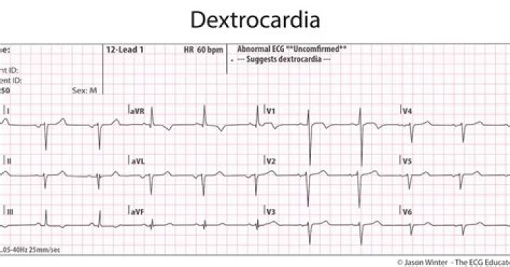
Most cases of dextrocardia are diagnosed using an electrocardiogram (EKG) and chest X-ray. An EKG that shows inverted or reversed electrical waves usually points to dextrocardia.Can a person be born with their heart on the right side?
Dextrocardia is a condition in which the heart is pointed toward the right side of the chest. Normally, the heart points toward the left. The condition is present at birth (congenital).
How many leads does a 12-lead ECG have?
Although it is called a 12-lead ECG, it uses only 10 electrodes. Certain electrodes are part of two pairs and thus provide two leads. Electrodes typically are self-adhesive pads with a conducting gel in the centre.
What is precordial leads in ECG?
Parts of an ECG The other six leads are considered “precordial leads” because they are placed on the torso (precordium). The six limb leads are called lead I, II, III, aVL, aVR and aVF. The letter “a” stands for “augmented,” as these leads are calculated as a combination of leads I, II and III.
What happens if ECG leads are put on incorrectly?
The analysis of ECG signals recorded from misplaced electrodes can lead to misinterpretation or even to significant diagnostic errors like incorrect recognition of anterior infarction, anteroseptal infarction, ventricular hypertrophy [9, 14], false diagnosis of ischemia, or Brugada syndrome [16, 24].What is Brugada syndrome?
Brugada (brew-GAH-dah) syndrome is a rare, but potentially life-threatening heart rhythm disorder that is sometimes inherited. People with Brugada syndrome have an increased risk of having irregular heart rhythms beginning in the lower chambers of the heart (ventricles).
What is the procedure for an ECG?Electrodes (small, plastic patches that stick to the skin) are placed at certain spots on the chest, arms, and legs. The electrodes are connected to an ECG machine by lead wires. The electrical activity of the heart is then measured, interpreted, and printed out. No electricity is sent into the body.
Article first time published onWhat is an electrocardiogram and how is it performed?
An electrocardiogram (ECG or EKG) records the electrical signal from your heart to check for different heart conditions. Electrodes are placed on your chest to record your heart’s electrical signals, which cause your heart to beat. The signals are shown as waves on an attached computer monitor or printer.
What should I do if my right side of my chest hurts?
Call 911 or seek emergency care if the pain or pressure in the right side of your chest is accompanied by: Sudden, severe abdominal pain. A rigid or tender abdomen. Vomiting of blood.
What is right sided heart failure?
Right-sided heart failure means your heart’s right ventricle is too weak to pump enough blood to the lungs. As a result: Blood builds up in your veins, vessels that carry blood from the body back to the heart. This buildup increases pressure in your veins.
What is it called when your organs are reversed?
Situs inversus is a genetic condition in which the organs in the chest and abdomen are positioned in a mirror image from their normal positions.
Is right-sided heart failure systolic or diastolic?
If you have systolic heart failure, it means your heart isn’t contracting well during heartbeats. If you have diastolic heart failure, it means your heart isn’t able to relax normally between beats. Both types of left-sided heart failure can lead to right-sided heart failure.
Which of the following symptoms might a client with right-sided heart failure exhibit *?
1 Symptoms of right-sided heart failure, such as dyspnea (shortness of breath), edema (swelling of the limbs), and fatigue can be severe.
Is right-sided heart failure reversible?
There is no cure for heart failure, but there are treatments for its symptoms. Talk to your doctor. They may suggest medications to make you more comfortable. In some cases, a procedure or surgery may be necessary.
Can you have dextrocardia without inversus?
In dextrocardia, the heart is on the right side of the thorax with or without situs inversus. When the heart is right sided with inverted atria, the stomach is right sided, and the liver is left sided, the combination is dextrocardia with situs inversus.
What is the difference between dextrocardia and Dextroposition?
Dextroposition describes a heart on the right with an apex to the left, secondary to extracardiac causes (right lung hypoplasia, pneumonectomy or diaphragmatic hernia). In contrast, dextrocardia results from cardiac chamber disarrangement.
Is dextrocardia a heart disease?
Dextrocardia is a heart condition that makes the heart move out of its usual position. It points towards the right side of your chest instead of the left side. The condition is congenital, meaning that people are born with it, but it’s rare.
Are precordial leads bipolar?
For a routine analysis of the heart’s electrical activity an ECG recorded from 12 separate leads is used. A 12-lead ECG consists of three bipolar limb leads (I, II, and III), the unipolar limb leads (AVR, AVL, and AVF), and six unipolar chest leads, also called precordial or V leads, ( , , , , , and ).
How does ischemia present on ECG?
The most common ECG sign of myocardial ischemia is flat or down-sloping ST-segment depression of 1.0 mm or greater. This report draws attention to other much less common, but possibly equally important, ECG manifestations of myocardial ischemia.
Why are precordial leads called V leads?
These leads were originally constructed by Goldberger. In these leads the exploring electrode is compared with a reference which is based on an average of the other two limb electrodes. The letter a stands for augmented, V for voltage and R is right arm, L is left arm and F is foot.
What does aVR stand for in ECG?
LabelMeaning of labelPosition of lead on bodyAVrAugmented vector rightRight wristAVLAugmented vector leftLeft wristAVfAugmented vector footLeft foot
What is LQTS syndrome?
Long QT syndrome (LQTS) is an abnormal feature of the heart’s electrical system that can lead to a potentially life-threatening arrhythmia called torsades de pointes (pronounced torsad de pwant). Torsades de pointes may result in syncope (fainting) or sudden cardiac death.
What does Brugada look like on EKG?
Brugada syndrome is a disorder characterized by sudden death associated with one of several electrocardiographic (ECG) patterns characterized by incomplete right bundle-branch block and ST elevations in the anterior precordial leads.
What is an Ajmaline test?
Your doctor has recommended that you have an ajmaline challenge. The purpose of this test is to see if you are likely to have Brugada syndrome, which is a disorder that affects the cells of the heart, causing it to have an altered rhythm. Ajmaline is a drug which will show up ECG changes if you have Brugada syndrome.
What percentage of recorded EKGS are performed with incorrect placement of the leads?
You are reading an ECG outside of a hospital. Even though nearly 4 percent of all ECG’s recorded are done so with incorrect placement of the leads, it happens even more often before the patient even reaches the hospital.
Why is the right leg grounded in ECG?
The right leg electrode acts to reduce interference, and can be placed anywhere without an effect on the ECG results. Each lead measures the electric field created by the heart during the depolarization and repolarization of myocytes.
What does lead aVR look at?
The lead aVR is oriented to ‘look’ at the right upper side of the heart, and can provide specific information about the right ventricle outflow tract and basal part of the septum (10).
What can be detected in ECG?
- arrhythmias – where the heart beats too slowly, too quickly, or irregularly.
- coronary heart disease – where the heart’s blood supply is blocked or interrupted by a build-up of fatty substances.
- heart attacks – where the supply of blood to the heart is suddenly blocked.